6GPZ

Cell division regulator GpsB in complex with peptide fragment of L. monocytogenes Penicillin Binding Protein PBPA1


Experimental Data Snapshot

  • Method: X-RAY DIFFRACTION
  • Resolution: 1.60 Å
  • R-Value Free: 0.204 
  • R-Value Work: 0.173 
  • R-Value Observed: 0.175 

wwPDB Validation   3D Report Full Report


This is version 1.2 of the entry. See complete history


Literature

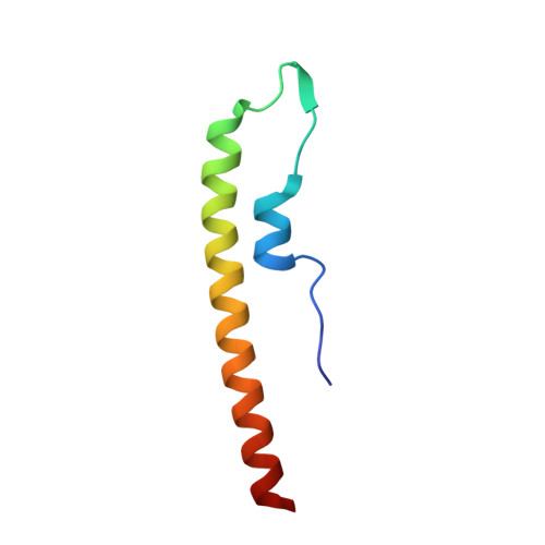
The cell cycle regulator GpsB functions as cytosolic adaptor for multiple cell wall enzymes.

Cleverley, R.M.Rutter, Z.J.Rismondo, J.Corona, F.Tsui, H.T.Alatawi, F.A.Daniel, R.A.Halbedel, S.Massidda, O.Winkler, M.E.Lewis, R.J.

(2019) Nat Commun 10: 261-261

  • DOI: https://doi.org/10.1038/s41467-018-08056-2
  • Primary Citation of Related Structures:  
    6GP7, 6GPZ, 6GQA, 6GQN

  • PubMed Abstract: 

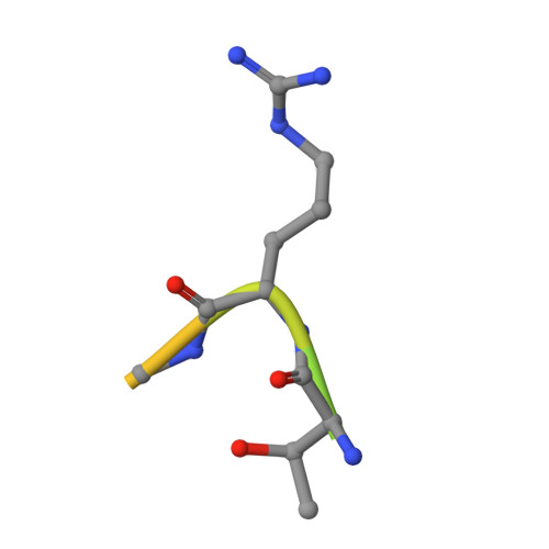
    Bacterial growth and cell division requires precise spatiotemporal regulation of the synthesis and remodelling of the peptidoglycan layer that surrounds the cytoplasmic membrane. GpsB is a cytosolic protein that affects cell wall synthesis by binding cytoplasmic mini-domains of peptidoglycan synthases to ensure their correct subcellular localisation. Here, we describe critical structural features for the interaction of GpsB with peptidoglycan synthases from three bacterial species (Bacillus subtilis, Listeria monocytogenes and Streptococcus pneumoniae) and suggest their importance for cell wall growth and viability in L. monocytogenes and S. pneumoniae. We use these structural motifs to identify novel partners of GpsB in B. subtilis and extend the members of the GpsB interactome in all three bacterial species. Our results support that GpsB functions as an adaptor protein that mediates the interaction between membrane proteins, scaffolding proteins, signalling proteins and enzymes to generate larger protein complexes at specific sites in a bacterial cell cycle-dependent manner.


  • Organizational Affiliation

    Institute for Cell and Molecular Biosciences, University of Newcastle, Newcastle upon Tyne, NE2 4HH, UK.


Macromolecules
Find similar proteins by:  (by identity cutoff)  |  3D Structure
Entity ID: 1
MoleculeChains Sequence LengthOrganismDetailsImage
Cell cycle protein GpsBA [auth B],
B [auth A]
63Bacillus subtilis subsp. subtilis str. 168Mutation(s): 1 
Gene Names: gpsBypsBBSU22180
UniProt
Find proteins for P0CI74 (Bacillus subtilis (strain 168))
Explore P0CI74 
Go to UniProtKB:  P0CI74
Entity Groups  
Sequence Clusters30% Identity50% Identity70% Identity90% Identity95% Identity100% Identity
UniProt GroupP0CI74
Sequence Annotations
Expand
  • Reference Sequence

Find similar proteins by:  Sequence   |   3D Structure  

Entity ID: 2
MoleculeChains Sequence LengthOrganismDetailsImage
LmPBPA1C [auth E]15Listeria monocytogenesMutation(s): 0 
Entity Groups  
Sequence Clusters30% Identity50% Identity70% Identity90% Identity95% Identity100% Identity
Sequence Annotations
Expand
  • Reference Sequence
Experimental Data & Validation

Experimental Data

  • Method: X-RAY DIFFRACTION
  • Resolution: 1.60 Å
  • R-Value Free: 0.204 
  • R-Value Work: 0.173 
  • R-Value Observed: 0.175 
  • Space Group: P 1 21 1
Unit Cell:
Length ( Å )Angle ( ˚ )
a = 26.577α = 90
b = 31.415β = 92.41
c = 81.032γ = 90
Software Package:
Software NamePurpose
PHENIXrefinement
DIALSdata reduction
Aimlessdata scaling
PHASERphasing

Structure Validation

View Full Validation Report


Entry History & Funding Information

Deposition Data


Funding OrganizationLocationGrant Number
Biotechnology and Biological Sciences Research CouncilUnited KingdomBB/M001180/1

Revision History  (Full details and data files)

  • Version 1.0: 2019-01-23
    Type: Initial release
  • Version 1.1: 2019-01-30
    Changes: Data collection, Database references
  • Version 1.2: 2024-01-17
    Changes: Data collection, Database references, Derived calculations, Refinement description