6GIZ

PURPLE ACID PHYTASE FROM WHEAT ISOFORM B2 - SUBSTRATE COMPLEX


Experimental Data Snapshot

  • Method: X-RAY DIFFRACTION
  • Resolution: 1.54 Å
  • R-Value Free: 0.167 
  • R-Value Work: 0.136 
  • R-Value Observed: 0.138 

wwPDB Validation   3D Report Full Report


Ligand Structure Quality Assessment 


This is version 2.2 of the entry. See complete history


Literature

Structure of a cereal purple acid phytase provides new insights to phytate degradation in plants.

Faba-Rodriguez, R.Gu, Y.Salmon, M.Dionisio, G.Brinch-Pedersen, H.Brearley, C.A.Hemmings, A.M.

(2022) Plant Commun 3: 100305-100305

  • DOI: https://doi.org/10.1016/j.xplc.2022.100305
  • Primary Citation of Related Structures:  
    6GIT, 6GIZ, 6GJ2, 6GJA

  • PubMed Abstract: 

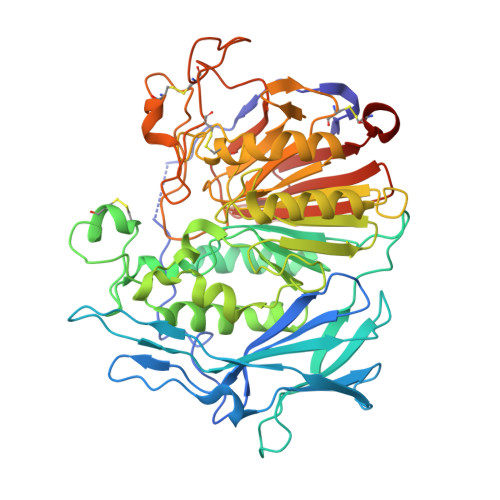
    Grain phytate, a mixed metal ion salt of inositol hexakisphosphate, accounts for 60%-80% of stored phosphorus in plants and is a potent antinutrient of non-ruminant animals including humans. Through neofunctionalization of purple acid phytases (PAPhy), some cereals such as wheat and rye have acquired particularly high mature grain phytase activity. As PAPhy activity supplies phosphate, liberates metal ions necessary for seedling emergence, and obviates antinutrient effects of phytate, its manipulation and control are targeted crop traits. Here we show the X-ray crystal structure of the b2 isoform of wheat PAPhy induced during germination. This high-resolution crystal structure suggests a model for phytate recognition that, validated by molecular dynamics simulations, implicates elements of two sequence inserts (termed PAPhy motifs) relative to a canonical metallophosphoesterase (MPE) domain in forming phytate-specific substrate specificity pockets. These motifs are well conserved in PAPhys from monocot cereals, enzymes which are characterized by high specificity for phytate. Tested by mutagenesis, residues His229 in PAPhy motif 4 and Lys410 in the MPE domain, both conserved in PAPhys, are found to strongly influence phytase activity. These results explain the observed phytase activity of cereal PAPhys and open the way to the rational engineering of phytase activity in planta .


  • Organizational Affiliation

    School of Chemistry, University of East Anglia, Norwich Research Park, Norwich NR4 7TJ, UK.


Macromolecules
Find similar proteins by:  (by identity cutoff)  |  3D Structure
Entity ID: 1
MoleculeChains Sequence LengthOrganismDetailsImage
Purple acid phosphatase516Triticum aestivumMutation(s): 0 
Gene Names: PAPhy
EC: 3.1.3.2
UniProt
Find proteins for F6MIW5 (Triticum aestivum)
Explore F6MIW5 
Go to UniProtKB:  F6MIW5
Entity Groups  
Sequence Clusters30% Identity50% Identity70% Identity90% Identity95% Identity100% Identity
UniProt GroupF6MIW5
Sequence Annotations
Expand
  • Reference Sequence
Oligosaccharides

Help

Entity ID: 2
MoleculeChains Length2D Diagram Glycosylation3D Interactions
2-acetamido-2-deoxy-beta-D-glucopyranose-(1-4)-2-acetamido-2-deoxy-beta-D-glucopyranose
B, C
2N-Glycosylation
Glycosylation Resources
GlyTouCan:  G42666HT
GlyCosmos:  G42666HT
GlyGen:  G42666HT
Small Molecules
Ligands 6 Unique
IDChains Name / Formula / InChI Key2D Diagram3D Interactions
NAG
Query on NAG

Download Ideal Coordinates CCD File 
F [auth A],
G [auth A],
H [auth A],
I [auth A],
J [auth A]
2-acetamido-2-deoxy-beta-D-glucopyranose
C8 H15 N O6
OVRNDRQMDRJTHS-FMDGEEDCSA-N
PGE
Query on PGE

Download Ideal Coordinates CCD File 
N [auth A],
O [auth A],
P [auth A]
TRIETHYLENE GLYCOL
C6 H14 O4
ZIBGPFATKBEMQZ-UHFFFAOYSA-N
PEG
Query on PEG

Download Ideal Coordinates CCD File 
Q [auth A],
R [auth A],
S [auth A],
T [auth A]
DI(HYDROXYETHYL)ETHER
C4 H10 O3
MTHSVFCYNBDYFN-UHFFFAOYSA-N
PO4
Query on PO4

Download Ideal Coordinates CCD File 
K [auth A],
L [auth A],
M [auth A]
PHOSPHATE ION
O4 P
NBIIXXVUZAFLBC-UHFFFAOYSA-K
EDO
Query on EDO

Download Ideal Coordinates CCD File 
AA [auth A]
BA [auth A]
U [auth A]
V [auth A]
W [auth A]
AA [auth A],
BA [auth A],
U [auth A],
V [auth A],
W [auth A],
X [auth A],
Y [auth A],
Z [auth A]
1,2-ETHANEDIOL
C2 H6 O2
LYCAIKOWRPUZTN-UHFFFAOYSA-N
FE
Query on FE

Download Ideal Coordinates CCD File 
D [auth A],
E [auth A]
FE (III) ION
Fe
VTLYFUHAOXGGBS-UHFFFAOYSA-N
Experimental Data & Validation

Experimental Data

  • Method: X-RAY DIFFRACTION
  • Resolution: 1.54 Å
  • R-Value Free: 0.167 
  • R-Value Work: 0.136 
  • R-Value Observed: 0.138 
  • Space Group: H 3
Unit Cell:
Length ( Å )Angle ( ˚ )
a = 126.73α = 90
b = 126.73β = 90
c = 107.04γ = 120
Software Package:
Software NamePurpose
Aimlessdata scaling
PHENIXrefinement
PDB_EXTRACTdata extraction
XDSdata reduction
PHASERphasing

Structure Validation

View Full Validation Report


Ligand Structure Quality Assessment 


Entry History & Funding Information

Deposition Data


Funding OrganizationLocationGrant Number
Biotechnology and Biological Sciences Research CouncilUnited KingdomBB/M022978/1

Revision History  (Full details and data files)

  • Version 1.0: 2019-11-27
    Type: Initial release
  • Version 2.0: 2020-07-29
    Type: Remediation
    Reason: Carbohydrate remediation
    Changes: Atomic model, Data collection, Derived calculations, Structure summary
  • Version 2.1: 2022-06-08
    Changes: Advisory, Database references, Structure summary
  • Version 2.2: 2024-01-17
    Changes: Data collection, Refinement description