6G1B

Corynebacterium glutamicum OxyR, oxidized form


Experimental Data Snapshot

  • Method: X-RAY DIFFRACTION
  • Resolution: 2.28 Å
  • R-Value Free: 0.230 
  • R-Value Work: 0.196 
  • R-Value Observed: 0.198 

wwPDB Validation   3D Report Full Report


Ligand Structure Quality Assessment 


This is version 1.1 of the entry. See complete history


Literature

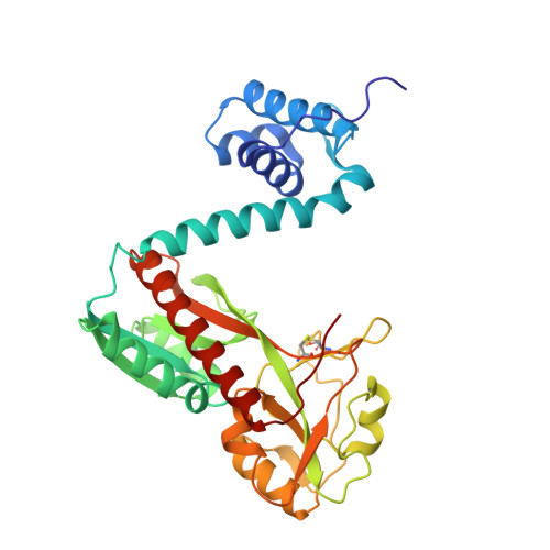
Structural snapshots of OxyR reveal the peroxidatic mechanism of H2O2sensing.

Pedre, B.Young, D.Charlier, D.Mourenza, A.Rosado, L.A.Marcos-Pascual, L.Wahni, K.Martens, E.G de la Rubia, A.Belousov, V.V.Mateos, L.M.Messens, J.

(2018) Proc Natl Acad Sci U S A 115: E11623-E11632

  • DOI: https://doi.org/10.1073/pnas.1807954115
  • Primary Citation of Related Structures:  
    6G1B, 6G1D, 6G4R

  • PubMed Abstract: 

    Hydrogen peroxide (H 2 O 2 ) is a strong oxidant capable of oxidizing cysteinyl thiolates, yet only a few cysteine-containing proteins have exceptional reactivity toward H 2 O 2 One such example is the prokaryotic transcription factor OxyR, which controls the antioxidant response in bacteria, and which specifically and rapidly reduces H 2 O 2 In this study, we present crystallographic evidence for the H 2 O 2 -sensing mechanism and H 2 O 2 -dependent structural transition of Corynebacterium glutamicum OxyR by capturing the reduced and H 2 O 2 -bound structures of a serine mutant of the peroxidatic cysteine, and the full-length crystal structure of disulfide-bonded oxidized OxyR. In the H 2 O 2 -bound structure, we pinpoint the key residues for the peroxidatic reduction of H 2 O 2 , and relate this to mutational assays showing that the conserved active-site residues T107 and R278 are critical for effective H 2 O 2 reduction. Furthermore, we propose an allosteric mode of structural change, whereby a localized conformational change arising from H 2 O 2 -induced intramolecular disulfide formation drives a structural shift at the dimerization interface of OxyR, leading to overall changes in quaternary structure and an altered DNA-binding topology and affinity at the catalase promoter region. This study provides molecular insights into the overall OxyR transcription mechanism regulated by H 2 O 2 .


  • Organizational Affiliation

    Center for Structural Biology, Vlaams Instituut voor Biotechnologie-Vrije Universiteit Brussel, B-1050 Brussels, Belgium.


Macromolecules
Find similar proteins by:  (by identity cutoff)  |  3D Structure
Entity ID: 1
MoleculeChains Sequence LengthOrganismDetailsImage
Hydrogen peroxide-inducible genes activatorA [auth J]327Corynebacterium glutamicumMutation(s): 1 
Gene Names: C0I99_13405
UniProt
Find proteins for Q8NP91 (Corynebacterium glutamicum (strain ATCC 13032 / DSM 20300 / BCRC 11384 / JCM 1318 / LMG 3730 / NCIMB 10025))
Explore Q8NP91 
Go to UniProtKB:  Q8NP91
Entity Groups  
Sequence Clusters30% Identity50% Identity70% Identity90% Identity95% Identity100% Identity
UniProt GroupQ8NP91
Sequence Annotations
Expand
  • Reference Sequence
Find similar proteins by:  (by identity cutoff)  |  3D Structure
Entity ID: 2
MoleculeChains Sequence LengthOrganismDetailsImage
Hydrogen peroxide-inducible genes activator326Corynebacterium glutamicumMutation(s): 1 
Gene Names: C0I99_13405
UniProt
Find proteins for Q8NP91 (Corynebacterium glutamicum (strain ATCC 13032 / DSM 20300 / BCRC 11384 / JCM 1318 / LMG 3730 / NCIMB 10025))
Explore Q8NP91 
Go to UniProtKB:  Q8NP91
Entity Groups  
Sequence Clusters30% Identity50% Identity70% Identity90% Identity95% Identity100% Identity
UniProt GroupQ8NP91
Sequence Annotations
Expand
  • Reference Sequence
Small Molecules
Ligands 5 Unique
IDChains Name / Formula / InChI Key2D Diagram3D Interactions
1PS
Query on 1PS

Download Ideal Coordinates CCD File 
E [auth J]3-PYRIDINIUM-1-YLPROPANE-1-SULFONATE
C8 H11 N O3 S
REEBJQTUIJTGAL-UHFFFAOYSA-N
NI
Query on NI

Download Ideal Coordinates CCD File 
C [auth J]NICKEL (II) ION
Ni
VEQPNABPJHWNSG-UHFFFAOYSA-N
FMT
Query on FMT

Download Ideal Coordinates CCD File 
D [auth J],
I [auth B],
J [auth B]
FORMIC ACID
C H2 O2
BDAGIHXWWSANSR-UHFFFAOYSA-N
PEO
Query on PEO

Download Ideal Coordinates CCD File 
G [auth J],
H [auth J],
K [auth B]
HYDROGEN PEROXIDE
H2 O2
MHAJPDPJQMAIIY-UHFFFAOYSA-N
NA
Query on NA

Download Ideal Coordinates CCD File 
F [auth J]SODIUM ION
Na
FKNQFGJONOIPTF-UHFFFAOYSA-N
Modified Residues  1 Unique
IDChains TypeFormula2D DiagramParent
SME
Query on SME
A [auth J]L-PEPTIDE LINKINGC5 H11 N O3 SMET
Experimental Data & Validation

Experimental Data

  • Method: X-RAY DIFFRACTION
  • Resolution: 2.28 Å
  • R-Value Free: 0.230 
  • R-Value Work: 0.196 
  • R-Value Observed: 0.198 
  • Space Group: C 1 2 1
Unit Cell:
Length ( Å )Angle ( ˚ )
a = 161.307α = 90
b = 46.186β = 121.92
c = 116.692γ = 90
Software Package:
Software NamePurpose
XDSdata reduction
Aimlessdata scaling
REFMACrefinement
PDB_EXTRACTdata extraction
PHASERphasing

Structure Validation

View Full Validation Report


Ligand Structure Quality Assessment 


Entry History & Funding Information

Deposition Data


Funding OrganizationLocationGrant Number
VIBBelgium--
IWTBelgium--
HerculesBelgiumHERC16
FWOBelgiumG.0D79.14N
VUB Strategic Research ProgrammeBelgiumSRP34
Junta de Castilla y LeonSpainLE326U14
University of LeonSpainUXXI2016/00127

Revision History  (Full details and data files)

  • Version 1.0: 2018-12-05
    Type: Initial release
  • Version 1.1: 2018-12-19
    Changes: Data collection, Database references