6FXP

Crystal structure of S. aureus glucosaminidase B


Experimental Data Snapshot

  • Method: X-RAY DIFFRACTION
  • Resolution: 2.03 Å
  • R-Value Free: 0.220 
  • R-Value Work: 0.212 
  • R-Value Observed: 0.212 

wwPDB Validation   3D Report Full Report


This is version 1.1 of the entry. See complete history


Literature

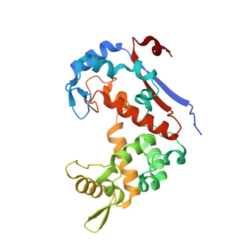
Domain sliding of two Staphylococcus aureus N-acetylglucosaminidases enables their substrate-binding prior to its catalysis.

Pintar, S.Borisek, J.Usenik, A.Perdih, A.Turk, D.

(2020) Commun Biol 3: 178-178

  • DOI: https://doi.org/10.1038/s42003-020-0911-7
  • Primary Citation of Related Structures:  
    6FXO, 6FXP

  • PubMed Abstract: 

    To achieve productive binding, enzymes and substrates must align their geometries to complement each other along an entire substrate binding site, which may require enzyme flexibility. In pursuit of novel drug targets for the human pathogen S. aureus, we studied peptidoglycan N-acetylglucosaminidases, whose structures are composed of two domains forming a V-shaped active site cleft. Combined insights from crystal structures supported by site-directed mutagenesis, modeling, and molecular dynamics enabled us to elucidate the substrate binding mechanism of SagB and AtlA-gl. This mechanism requires domain sliding from the open form observed in their crystal structures, leading to polysaccharide substrate binding in the closed form, which can enzymatically process the bound substrate. We suggest that these two hydrolases must exhibit unusual extents of flexibility to cleave the rigid structure of a bacterial cell wall.


  • Organizational Affiliation

    Department of Biochemistry, Molecular and Structural Biology, Jožef Stefan Institute, Jamova cesta 39, 1000, Ljubljana, Slovenia.


Macromolecules
Find similar proteins by:  (by identity cutoff)  |  3D Structure
Entity ID: 1
MoleculeChains Sequence LengthOrganismDetailsImage
Uncharacterized protein
A, B
254Staphylococcus aureus subsp. aureus Mu50Mutation(s): 0 
Gene Names: SAV1775
UniProt
Find proteins for A0A0H3JSV1 (Staphylococcus aureus (strain Mu50 / ATCC 700699))
Explore A0A0H3JSV1 
Go to UniProtKB:  A0A0H3JSV1
Entity Groups  
Sequence Clusters30% Identity50% Identity70% Identity90% Identity95% Identity100% Identity
UniProt GroupA0A0H3JSV1
Sequence Annotations
Expand
  • Reference Sequence
Small Molecules
Ligands 3 Unique
IDChains Name / Formula / InChI Key2D Diagram3D Interactions
PG4
Query on PG4

Download Ideal Coordinates CCD File 
C [auth A]
D [auth A]
G [auth A]
H [auth A]
I [auth A]
C [auth A],
D [auth A],
G [auth A],
H [auth A],
I [auth A],
J [auth B],
L [auth B],
M [auth B]
TETRAETHYLENE GLYCOL
C8 H18 O5
UWHCKJMYHZGTIT-UHFFFAOYSA-N
CL
Query on CL

Download Ideal Coordinates CCD File 
E [auth A],
K [auth B]
CHLORIDE ION
Cl
VEXZGXHMUGYJMC-UHFFFAOYSA-M
NA
Query on NA

Download Ideal Coordinates CCD File 
F [auth A]SODIUM ION
Na
FKNQFGJONOIPTF-UHFFFAOYSA-N
Experimental Data & Validation

Experimental Data

  • Method: X-RAY DIFFRACTION
  • Resolution: 2.03 Å
  • R-Value Free: 0.220 
  • R-Value Work: 0.212 
  • R-Value Observed: 0.212 
  • Space Group: P 61 2 2
Unit Cell:
Length ( Å )Angle ( ˚ )
a = 151.77α = 90
b = 151.77β = 90
c = 122.51γ = 120
Software Package:
Software NamePurpose
MAINrefinement
XDSdata reduction
XDSdata scaling
SHELXDEphasing

Structure Validation

View Full Validation Report


Entry History & Funding Information

Deposition Data


Funding OrganizationLocationGrant Number
ARRSSloveniaP1-0048

Revision History  (Full details and data files)

  • Version 1.0: 2019-03-20
    Type: Initial release
  • Version 1.1: 2020-09-30
    Changes: Database references, Derived calculations