6FXJ

Structure of coproheme decarboxylase from Listeria monocytogenes in complex with iron coproporphyrin III


Experimental Data Snapshot

  • Method: X-RAY DIFFRACTION
  • Resolution: 1.79 Å
  • R-Value Free: 0.214 
  • R-Value Work: 0.186 
  • R-Value Observed: 0.187 

wwPDB Validation   3D Report Full Report


Ligand Structure Quality Assessment 


This is version 1.2 of the entry. See complete history


Literature

Redox Cofactor Rotates during Its Stepwise Decarboxylation: Molecular Mechanism of Conversion of Coproheme to Hemeb.

Milazzo, L.Gabler, T.Puhringer, D.Jandova, Z.Maresch, D.Michlits, H.Pfanzagl, V.Djinovic-Carugo, K.Oostenbrink, C.Furtmuller, P.G.Obinger, C.Smulevich, G.Hofbauer, S.

(2019) ACS Catal 9: 6766-6782

  • DOI: https://doi.org/10.1021/acscatal.9b00963
  • Primary Citation of Related Structures:  
    6FXJ, 6FXQ

  • PubMed Abstract: 

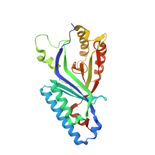
    Coproheme decarboxylase (ChdC) catalyzes the last step in the heme biosynthesis pathway of monoderm bacteria with coproheme acting both as redox cofactor and substrate. Hydrogen peroxide mediates the stepwise decarboxylation of propionates 2 and 4 of coproheme. Here we present the crystal structures of coproheme-loaded ChdC from Listeria monocytogenes (LmChdC) and the three-propionate intermediate, for which the propionate at position 2 (p2) has been converted to a vinyl group and is rotated by 90° compared to the coproheme complex structure. Single, double, and triple mutants of LmChdC, in which H-bonding interactions to propionates 2, 4, 6, and 7 were eliminated, allowed us to obtain the assignment of the coproheme propionates by resonance Raman spectroscopy and to follow the H 2 O 2 -mediated conversion of coproheme to heme b . Substitution of H 2 O 2 by chlorite allowed us to monitor compound I formation in the inactive Y147H variant which lacks the catalytically essential Y147. This residue was demonstrated to be oxidized during turnover by using the spin-trap 2-methyl-2-nitrosopropane. Based on these findings and the data derived from molecular dynamics simulations of cofactor structures in distinct poses, we propose a reaction mechanism for the stepwise decarboxylation of coproheme that includes a 90° rotation of the intermediate three-propionate redox cofactor.


  • Organizational Affiliation

    Dipartimento di Chimica "Ugo Schiff", Università di Firenze, Via della Lastruccia 3-13, I-50019 Sesto Fiorentino (FI), Italy.


Macromolecules
Find similar proteins by:  (by identity cutoff)  |  3D Structure
Entity ID: 1
MoleculeChains Sequence LengthOrganismDetailsImage
Putative heme-dependent peroxidase lmo2113
A, B, C, D, E
251Listeria monocytogenes EGD-eMutation(s): 0 
Gene Names: lmo2113
EC: 1.11.1
UniProt
Find proteins for Q8Y5F1 (Listeria monocytogenes serovar 1/2a (strain ATCC BAA-679 / EGD-e))
Explore Q8Y5F1 
Go to UniProtKB:  Q8Y5F1
Entity Groups  
Sequence Clusters30% Identity50% Identity70% Identity90% Identity95% Identity100% Identity
UniProt GroupQ8Y5F1
Sequence Annotations
Expand
  • Reference Sequence
Small Molecules
Ligands 4 Unique
IDChains Name / Formula / InChI Key2D Diagram3D Interactions
FEC (Subject of Investigation/LOI)
Query on FEC

Download Ideal Coordinates CCD File 
G [auth A],
J [auth B],
M [auth C],
S [auth D],
Z [auth E]
1,3,5,8-TETRAMETHYL-PORPHINE-2,4,6,7-TETRAPROPIONIC ACID FERROUS COMPLEX
C36 H36 Fe N4 O8
FEDZMOFKVKOYTI-RGGAHWMASA-L
POL
Query on POL

Download Ideal Coordinates CCD File 
AA [auth E]
BA [auth E]
K [auth B]
L [auth B]
N [auth C]
AA [auth E],
BA [auth E],
K [auth B],
L [auth B],
N [auth C],
O [auth C],
P [auth C],
T [auth D],
U [auth D],
V [auth D]
N-PROPANOL
C3 H8 O
BDERNNFJNOPAEC-UHFFFAOYSA-N
CL
Query on CL

Download Ideal Coordinates CCD File 
X [auth D]CHLORIDE ION
Cl
VEXZGXHMUGYJMC-UHFFFAOYSA-M
NA
Query on NA

Download Ideal Coordinates CCD File 
F [auth A]
H [auth A]
I [auth B]
Q [auth C]
R [auth D]
F [auth A],
H [auth A],
I [auth B],
Q [auth C],
R [auth D],
W [auth D],
Y [auth E]
SODIUM ION
Na
FKNQFGJONOIPTF-UHFFFAOYSA-N
Experimental Data & Validation

Experimental Data

  • Method: X-RAY DIFFRACTION
  • Resolution: 1.79 Å
  • R-Value Free: 0.214 
  • R-Value Work: 0.186 
  • R-Value Observed: 0.187 
  • Space Group: P 1 21 1
Unit Cell:
Length ( Å )Angle ( ˚ )
a = 77.87α = 90
b = 129.11β = 105.96
c = 77.92γ = 90
Software Package:
Software NamePurpose
PHENIXrefinement
PHENIXrefinement
XDSdata reduction
XSCALEdata scaling
PHASERphasing

Structure Validation

View Full Validation Report


Ligand Structure Quality Assessment 


Entry History & Funding Information

Deposition Data


Funding OrganizationLocationGrant Number
Austrian Science FundAustriaP29099

Revision History  (Full details and data files)

  • Version 1.0: 2019-07-10
    Type: Initial release
  • Version 1.1: 2019-08-28
    Changes: Data collection, Database references
  • Version 1.2: 2024-01-17
    Changes: Advisory, Data collection, Database references, Derived calculations, Refinement description