6FT9

Crystal structure of CLK1 in complex with inhibitor 16


Experimental Data Snapshot

  • Method: X-RAY DIFFRACTION
  • Resolution: 1.87 Å
  • R-Value Free: 0.202 
  • R-Value Work: 0.166 
  • R-Value Observed: 0.168 

wwPDB Validation   3D Report Full Report


Ligand Structure Quality Assessment 


This is version 1.1 of the entry. See complete history


Literature

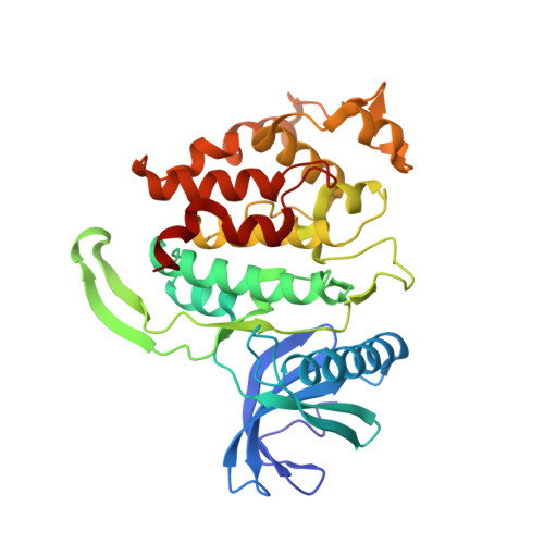
Molecular structures of cdc2-like kinases in complex with a new inhibitor chemotype.

Walter, A.Chaikuad, A.Helmer, R.Loaec, N.Preu, L.Ott, I.Knapp, S.Meijer, L.Kunick, C.

(2018) PLoS One 13: e0196761-e0196761

  • DOI: https://doi.org/10.1371/journal.pone.0196761
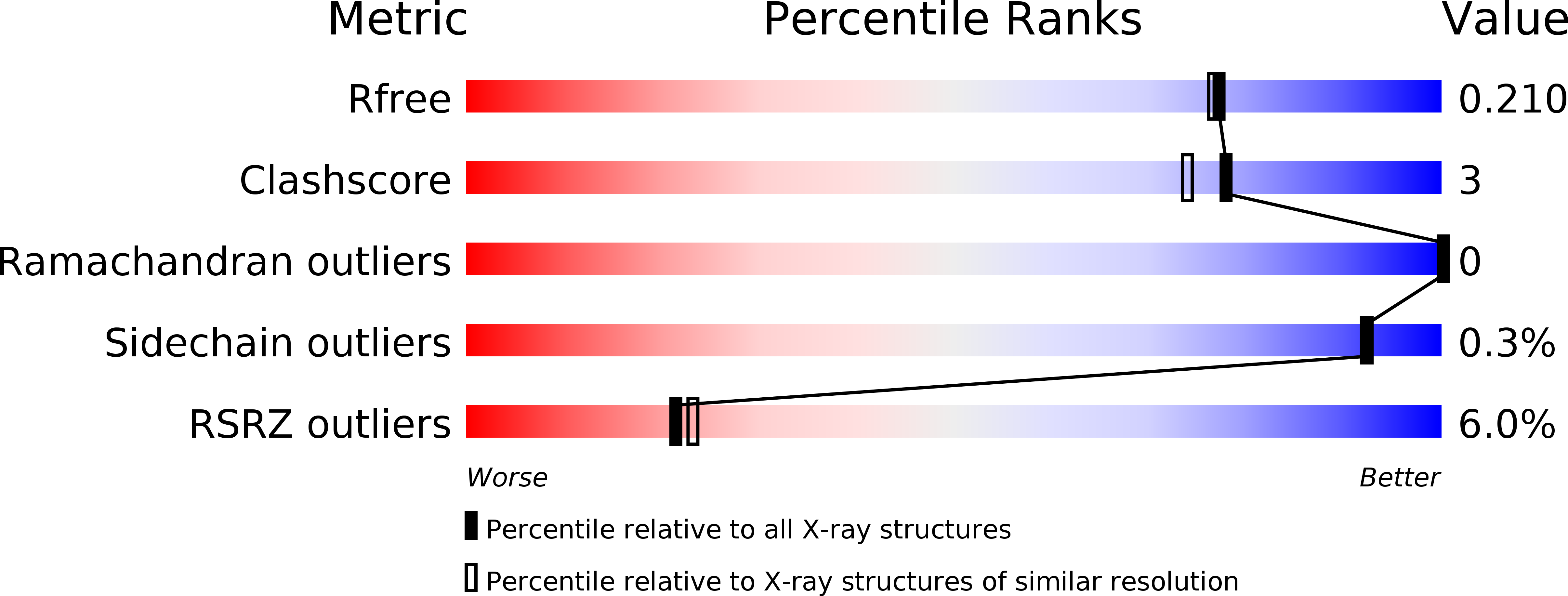
  • Primary Citation of Related Structures:  
    6FT7, 6FT8, 6FT9

  • PubMed Abstract: 

    Cdc2-like kinases (CLKs) represent a family of serine-threonine kinases involved in the regulation of splicing by phosphorylation of SR-proteins and other splicing factors. Although compounds acting against CLKs have been described, only a few show selectivity against dual-specificity tyrosine phosphorylation regulated-kinases (DYRKs). We here report a novel CLK inhibitor family based on a 6,7-dihydropyrrolo[3,4-g]indol-8(1H)-one core scaffold. Within the series, 3-(3-chlorophenyl)-6,7-dihydropyrrolo[3,4-g]indol-8(1H)-one (KuWal151) was identified as inhibitor of CLK1, CLK2 and CLK4 with a high selectivity margin towards DYRK kinases. The compound displayed a potent antiproliferative activity in an array of cultured cancer cell lines. The X-ray structure analyses of three members of the new compound class co-crystallized with CLK proteins corroborated a molecular binding mode predicted by docking studies.


  • Organizational Affiliation

    Institut für Medizinische und Pharmazeutische Chemie, Technische Universität Braunschweig, Braunschweig, Germany.


Macromolecules
Find similar proteins by:  (by identity cutoff)  |  3D Structure
Entity ID: 1
MoleculeChains Sequence LengthOrganismDetailsImage
Dual specificity protein kinase CLK1
A, B, C
339Homo sapiensMutation(s): 1 
Gene Names: CLK1CLK
EC: 2.7.12.1
UniProt & NIH Common Fund Data Resources
Find proteins for P49759 (Homo sapiens)
Explore P49759 
Go to UniProtKB:  P49759
PHAROS:  P49759
GTEx:  ENSG00000013441 
Entity Groups  
Sequence Clusters30% Identity50% Identity70% Identity90% Identity95% Identity100% Identity
UniProt GroupP49759
Sequence Annotations
Expand
  • Reference Sequence
Small Molecules
Ligands 5 Unique
IDChains Name / Formula / InChI Key2D Diagram3D Interactions
E6W
Query on E6W

Download Ideal Coordinates CCD File 
D [auth A],
K [auth B],
P [auth C]
2-bromanyl-3-phenyl-1~{H}-pyrrolo[3,4-g]indol-8-one
C16 H9 Br N2 O
VKVBXHZFKDKMDA-UHFFFAOYSA-N
PO4
Query on PO4

Download Ideal Coordinates CCD File 
F [auth A],
M [auth B],
R [auth C]
PHOSPHATE ION
O4 P
NBIIXXVUZAFLBC-UHFFFAOYSA-K
GOL
Query on GOL

Download Ideal Coordinates CCD File 
G [auth A],
H [auth A],
J [auth A],
N [auth B],
S [auth C]
GLYCEROL
C3 H8 O3
PEDCQBHIVMGVHV-UHFFFAOYSA-N
BR
Query on BR

Download Ideal Coordinates CCD File 
E [auth A],
L [auth B],
Q [auth C]
BROMIDE ION
Br
CPELXLSAUQHCOX-UHFFFAOYSA-M
NA
Query on NA

Download Ideal Coordinates CCD File 
I [auth A],
O [auth B],
T [auth C]
SODIUM ION
Na
FKNQFGJONOIPTF-UHFFFAOYSA-N
Experimental Data & Validation

Experimental Data

  • Method: X-RAY DIFFRACTION
  • Resolution: 1.87 Å
  • R-Value Free: 0.202 
  • R-Value Work: 0.166 
  • R-Value Observed: 0.168 
  • Space Group: P 1 21 1
Unit Cell:
Length ( Å )Angle ( ˚ )
a = 56.4α = 90
b = 116.301β = 98.98
c = 91.33γ = 90
Software Package:
Software NamePurpose
REFMACrefinement
MOSFLMdata reduction
SCALAdata scaling
PHASERphasing

Structure Validation

View Full Validation Report


Ligand Structure Quality Assessment 


Entry History 

Revision History  (Full details and data files)

  • Version 1.0: 2018-05-16
    Type: Initial release
  • Version 1.1: 2024-01-17
    Changes: Data collection, Database references, Refinement description