6EZ3

Structure of CDPS from Staphylococcus haemolyticus


Experimental Data Snapshot

  • Method: X-RAY DIFFRACTION
  • Resolution: 3.00 Å
  • R-Value Free: 0.263 
  • R-Value Work: 0.220 
  • R-Value Observed: 0.222 

wwPDB Validation   3D Report Full Report


This is version 1.0 of the entry. See complete history


Literature

Structural basis for partition of the cyclodipeptide synthases into two subfamilies.

Bourgeois, G.Seguin, J.Babin, M.Belin, P.Moutiez, M.Mechulam, Y.Gondry, M.Schmitt, E.

(2018) J Struct Biol 203: 17-26

  • DOI: https://doi.org/10.1016/j.jsb.2018.03.001
  • Primary Citation of Related Structures:  
    5MLP, 5MLQ, 5OCD, 6EZ3

  • PubMed Abstract: 

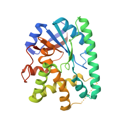
    Cyclodipeptide synthases (CDPSs) use two aminoacyl-tRNAs to catalyze the formation of two peptide bonds leading to cyclodipeptides that can be further used for the synthesis of diketopiperazines. It was shown that CDPSs fall into two subfamilies, NYH and XYP, characterized by the presence of specific sequence signatures. However, current understanding of CDPSs only comes from studies of enzymes from the NYH subfamily. The present study reveals the crystal structures of three CDPSs from the XYP subfamily. Comparison of the XYP and NYH enzymes shows that the two subfamilies mainly differ in the first half of their Rossmann fold. This gives a structural basis for the partition of CDPSs into two subfamilies. Despite these differences, the catalytic residues adopt similar positioning regardless of the subfamily suggesting that the XYP and NYH motifs correspond to two structural solutions to facilitate the reactivity of the catalytic serine residue.


  • Organizational Affiliation

    Laboratoire de Biochimie, Ecole polytechnique, CNRS, Université Paris-Saclay, 91128 Palaiseau cedex, France.


Macromolecules
Find similar proteins by:  (by identity cutoff)  |  3D Structure
Entity ID: 1
MoleculeChains Sequence LengthOrganismDetailsImage
Cyclo(L-leucyl-L-leucyl) synthase
A, B, C, D, E
A, B, C, D, E, F
234Staphylococcus haemolyticus JCSC1435Mutation(s): 0 
Gene Names: pSHaeC06
EC: 2.3.2.22
UniProt
Find proteins for Q4L2X9 (Staphylococcus haemolyticus (strain JCSC1435))
Explore Q4L2X9 
Go to UniProtKB:  Q4L2X9
Entity Groups  
Sequence Clusters30% Identity50% Identity70% Identity90% Identity95% Identity100% Identity
UniProt GroupQ4L2X9
Sequence Annotations
Expand
  • Reference Sequence
Small Molecules
Ligands 1 Unique
IDChains Name / Formula / InChI Key2D Diagram3D Interactions
SO4
Query on SO4

Download Ideal Coordinates CCD File 
G [auth A]
H [auth A]
I [auth B]
J [auth B]
K [auth C]
G [auth A],
H [auth A],
I [auth B],
J [auth B],
K [auth C],
L [auth C],
M [auth D],
N [auth D],
O [auth E],
P [auth F],
Q [auth F]
SULFATE ION
O4 S
QAOWNCQODCNURD-UHFFFAOYSA-L
Experimental Data & Validation

Experimental Data

  • Method: X-RAY DIFFRACTION
  • Resolution: 3.00 Å
  • R-Value Free: 0.263 
  • R-Value Work: 0.220 
  • R-Value Observed: 0.222 
  • Space Group: P 21 21 21
Unit Cell:
Length ( Å )Angle ( ˚ )
a = 46.381α = 90
b = 81.835β = 90
c = 384.026γ = 90
Software Package:
Software NamePurpose
BUSTERrefinement
XDSdata reduction
XDSdata scaling
PHASERphasing

Structure Validation

View Full Validation Report


Entry History & Funding Information

Deposition Data


Funding OrganizationLocationGrant Number
French National Research AgencyFrance--

Revision History  (Full details and data files)

  • Version 1.0: 2018-11-14
    Type: Initial release