6EWM

Crystal structure of heme free PORPHYROMONAS GINGIVALIS HEME-BINDING PROTEIN HMUY


Experimental Data Snapshot

  • Method: X-RAY DIFFRACTION
  • Resolution: 1.40 Å
  • R-Value Free: 0.171 
  • R-Value Work: 0.131 
  • R-Value Observed: 0.133 

wwPDB Validation   3D Report Full Report


This is version 1.2 of the entry. See complete history


Literature

Tannerella forsythiaTfo belongs toPorphyromonas gingivalisHmuY-like family of proteins but differs in heme-binding properties.

Bielecki, M.Antonyuk, S.Strange, R.W.Smalley, J.W.Mackiewicz, P.Smiga, M.Stepien, P.Olczak, M.Olczak, T.

(2018) Biosci Rep 38

  • DOI: https://doi.org/10.1042/BSR20181325
  • Primary Citation of Related Structures:  
    6EU8, 6EWM

  • PubMed Abstract: 

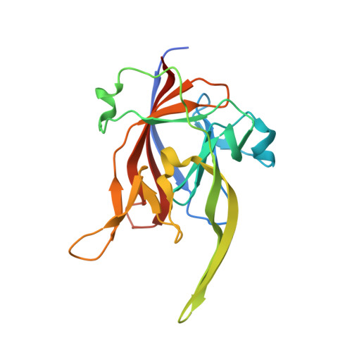
    Porphyromonas gingivalis is considered the principal etiologic agent and keystone pathogen of chronic periodontitis. As an auxotrophic bacterium, it must acquire heme to survive and multiply at the infection site. P. gingivalis HmuY is the first member of a novel family of hemophore-like proteins. Bacterial heme-binding proteins usually use histidine-methionine or histidine-tyrosine residues to ligate heme-iron, whereas P. gingivalis HmuY uses two histidine residues. We hypothesized that other 'red complex' members, i.e. Tannerella forsythia and Treponema denticola might utilize similar heme uptake mechanisms to the P. gingivalis HmuY. Comparative and phylogenetic analyses suggested differentiation of HmuY homologs and low conservation of heme-coordinating histidine residues present in HmuY. The homologs were subjected to duplication before divergence of Bacteroidetes lineages, which could facilitate evolution of functional diversification. We found that T. denticola does not code an HmuY homolog. T. forsythia protein, termed as Tfo, binds heme, but preferentially in the ferrous form, and sequesters heme from the albumin-heme complex under reducing conditions. In agreement with that, the 3D structure of Tfo differs from that of HmuY in the folding of heme-binding pocket, containing two methionine residues instead of two histidine residues coordinating heme in HmuY. Heme binding to apo-HmuY is accompanied by movement of the loop carrying the His 166 residue, closing the heme-binding pocket. Molecular dynamics simulations (MD) demonstrated that this conformational change also occurs in Tfo. In conclusion, our findings suggest that HmuY-like family might comprise proteins subjected during evolution to significant diversification, resulting in different heme-binding properties.


  • Organizational Affiliation

    Faculty of Biotechnology, University of Wrocław, Wrocław, Poland.


Macromolecules
Find similar proteins by:  (by identity cutoff)  |  3D Structure
Entity ID: 1
MoleculeChains Sequence LengthOrganismDetailsImage
Haemophore HmuYA,
B [auth F]
191Porphyromonas gingivalisMutation(s): 0 
Gene Names: hmuY'hmuYAT291_03715PGIN_13-1_01941PGIN_YH522_00859
UniProt
Find proteins for A2I2W2 (Porphyromonas gingivalis)
Explore A2I2W2 
Go to UniProtKB:  A2I2W2
Entity Groups  
Sequence Clusters30% Identity50% Identity70% Identity90% Identity95% Identity100% Identity
UniProt GroupA2I2W2
Sequence Annotations
Expand
  • Reference Sequence
Small Molecules
Ligands 3 Unique
IDChains Name / Formula / InChI Key2D Diagram3D Interactions
MES
Query on MES

Download Ideal Coordinates CCD File 
F [auth A]2-(N-MORPHOLINO)-ETHANESULFONIC ACID
C6 H13 N O4 S
SXGZJKUKBWWHRA-UHFFFAOYSA-N
SO4
Query on SO4

Download Ideal Coordinates CCD File 
C [auth A]
D [auth A]
E [auth A]
I [auth F]
J [auth F]
C [auth A],
D [auth A],
E [auth A],
I [auth F],
J [auth F],
K [auth F]
SULFATE ION
O4 S
QAOWNCQODCNURD-UHFFFAOYSA-L
GOL
Query on GOL

Download Ideal Coordinates CCD File 
G [auth A],
H [auth A],
L [auth F],
M [auth F],
N [auth F]
GLYCEROL
C3 H8 O3
PEDCQBHIVMGVHV-UHFFFAOYSA-N
Experimental Data & Validation

Experimental Data

  • Method: X-RAY DIFFRACTION
  • Resolution: 1.40 Å
  • R-Value Free: 0.171 
  • R-Value Work: 0.131 
  • R-Value Observed: 0.133 
  • Space Group: P 1 21 1
Unit Cell:
Length ( Å )Angle ( ˚ )
a = 59.334α = 90
b = 42.947β = 107.87
c = 95.075γ = 90
Software Package:
Software NamePurpose
REFMACrefinement
DIALSdata reduction
Aimlessdata scaling
MOLREPphasing

Structure Validation

View Full Validation Report


Entry History & Funding Information

Deposition Data


Funding OrganizationLocationGrant Number
National Science CenterPoland2015/17/B/NZ6/01969
European Regional Development FundPolandPOIG 01.01.02-02-003/08/00

Revision History  (Full details and data files)

  • Version 1.0: 2018-10-24
    Type: Initial release
  • Version 1.1: 2018-11-07
    Changes: Data collection, Database references
  • Version 1.2: 2024-01-17
    Changes: Data collection, Database references, Refinement description