6ENH

Crystal structure of the 43K ATPase domain of Thermus thermophilus gyrase B in complex with an aminocoumarin


Experimental Data Snapshot

  • Method: X-RAY DIFFRACTION
  • Resolution: 1.94 Å
  • R-Value Free: 0.233 
  • R-Value Work: 0.197 
  • R-Value Observed: 0.199 

wwPDB Validation   3D Report Full Report


Ligand Structure Quality Assessment 


This is version 1.2 of the entry. See complete history


Literature

Structural Basis for DNA Gyrase Interaction with Coumermycin A1.

Vanden Broeck, A.McEwen, A.G.Chebaro, Y.Potier, N.Lamour, V.

(2019) J Med Chem 62: 4225-4231

  • DOI: https://doi.org/10.1021/acs.jmedchem.8b01928
  • Primary Citation of Related Structures:  
    6ENG, 6ENH

  • PubMed Abstract: 

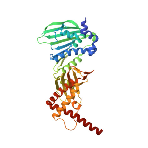
    Coumermycin A1 is a natural aminocoumarin that inhibits bacterial DNA gyrase, a member of the GHKL proteins superfamily. We report here the first cocrystal structures of gyrase B bound to coumermycin A1, revealing that one coumermycin A1 molecule traps simultaneously two ATP-binding sites. The inhibited dimers from different species adopt distinct sequence-dependent conformations, alternative to the ATP-bound form. These structures provide a basis for the rational development of coumermycin A1 derivatives for antibiotherapy and biotechnology applications.


  • Organizational Affiliation

    Integrated Structural Biology Department, IGBMC, UMR7104 CNRS, U1258 Inserm, University of Strasbourg, Illkirch 67404 , France.


Macromolecules
Find similar proteins by:  (by identity cutoff)  |  3D Structure
Entity ID: 1
MoleculeChains Sequence LengthOrganismDetailsImage
DNA gyrase subunit B393Thermus thermophilus HB8Mutation(s): 0 
Gene Names: gyrBTTHA1586
EC: 5.99.1.3
UniProt
Find proteins for Q5SHZ4 (Thermus thermophilus (strain ATCC 27634 / DSM 579 / HB8))
Explore Q5SHZ4 
Go to UniProtKB:  Q5SHZ4
Entity Groups  
Sequence Clusters30% Identity50% Identity70% Identity90% Identity95% Identity100% Identity
UniProt GroupQ5SHZ4
Sequence Annotations
Expand
  • Reference Sequence
Experimental Data & Validation

Experimental Data

  • Method: X-RAY DIFFRACTION
  • Resolution: 1.94 Å
  • R-Value Free: 0.233 
  • R-Value Work: 0.197 
  • R-Value Observed: 0.199 
  • Space Group: C 1 2 1
Unit Cell:
Length ( Å )Angle ( ˚ )
a = 166.37α = 90
b = 37.85β = 110.24
c = 66.75γ = 90
Software Package:
Software NamePurpose
BUSTERrefinement
XSCALEdata scaling
PDB_EXTRACTdata extraction
XDSdata reduction
PHASERphasing

Structure Validation

View Full Validation Report


Ligand Structure Quality Assessment 


Entry History & Funding Information

Deposition Data


Funding OrganizationLocationGrant Number
French National Research AgencyFranceANR-10-INSB-05-01
French National Research AgencyFranceANR-10-LABX-0030-INRT

Revision History  (Full details and data files)

  • Version 1.0: 2019-04-10
    Type: Initial release
  • Version 1.1: 2019-05-08
    Changes: Data collection, Database references
  • Version 1.2: 2024-01-17
    Changes: Data collection, Database references, Refinement description