6EDH

Taurine:2OG dioxygenase (TauD) bound to the vanadyl ion, taurine, and succinate


Experimental Data Snapshot

  • Method: X-RAY DIFFRACTION
  • Resolution: 1.73 Å
  • R-Value Free: 0.205 
  • R-Value Work: 0.183 
  • R-Value Observed: 0.183 

wwPDB Validation   3D Report Full Report


This is version 1.5 of the entry. See complete history


Literature

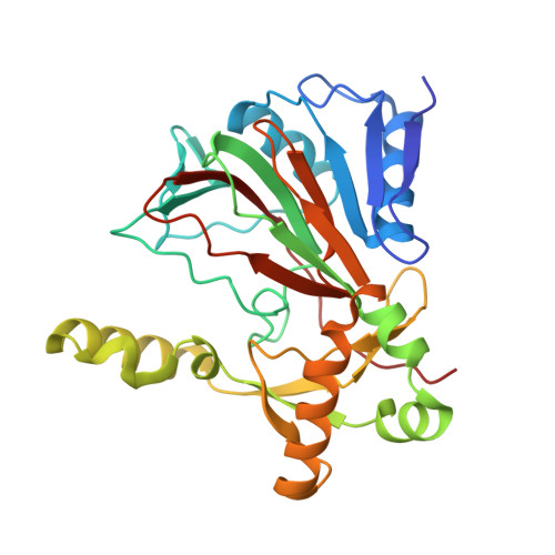
Structure of a Ferryl Mimic in the Archetypal Iron(II)- and 2-(Oxo)-glutarate-Dependent Dioxygenase, TauD.

Davis, K.M.Altmyer, M.Martinie, R.J.Schaperdoth, I.Krebs, C.Bollinger Jr., J.M.Boal, A.K.

(2019) Biochemistry 58: 4218-4223

  • DOI: https://doi.org/10.1021/acs.biochem.9b00598
  • Primary Citation of Related Structures:  
    6EDH

  • PubMed Abstract: 

    Iron(II)- and 2-(oxo)-glutarate-dependent (Fe/2OG) oxygenases catalyze a diverse array of oxidation reactions via a common iron(IV)-oxo (ferryl) intermediate. Although the intermediate has been characterized spectroscopically, its short lifetime has precluded crystallograhic characterization. In solution, the ferryl was first observed directly in the archetypal Fe/2OG hydroxylase, taurine:2OG dioxygenase (TauD). Here, we substitute the iron cofactor of TauD with the stable vanadium(IV)-oxo (vanadyl) ion to obtain crystal structures mimicking the key ferryl complex. Intriguingly, whereas the structure of the TauD·(V IV -oxo)·succinate·taurine complex exhibits the expected orientation of the V≡O bond- trans to the His255 ligand and toward the C-H bond to be cleaved, in what has been termed the in-line configuration-the TauD·(V IV -oxo) binary complex is best modeled with its oxo ligand trans to Asp101. This off-line-like configuration is similar to one recently posited as a means to avoid hydroxylation in Fe/2OG enzymes that direct other outcomes, though neither has been visualized in an Fe/2OG structure to date. Whereas an off-line ( trans to the proximal His) or off-line-like ( trans to the carboxylate ligand) ferryl is unlikely to be important in the hydroxylation reaction of TauD, the observation that the ferryl may deviate from an in-line orientation in the absence of the primary substrate may explain the enzyme's mysterious self-hydroxylation behavior, should the oxo ligand lie trans to His99. This finding reinforces the potential for analogous functional off-line oxo configurations in halogenases, desaturases, and/or cyclases.


Macromolecules
Find similar proteins by:  (by identity cutoff)  |  3D Structure
Entity ID: 1
MoleculeChains Sequence LengthOrganismDetailsImage
Alpha-ketoglutarate-dependent taurine dioxygenase
A, B
283Escherichia coli K-12Mutation(s): 0 
Gene Names: tauDssiDyaiGb0368JW0360
EC: 1.14.11.17
UniProt
Find proteins for P37610 (Escherichia coli (strain K12))
Explore P37610 
Go to UniProtKB:  P37610
Entity Groups  
Sequence Clusters30% Identity50% Identity70% Identity90% Identity95% Identity100% Identity
UniProt GroupP37610
Sequence Annotations
Expand
  • Reference Sequence
Small Molecules
Ligands 7 Unique
IDChains Name / Formula / InChI Key2D Diagram3D Interactions
1PE
Query on 1PE

Download Ideal Coordinates CCD File 
H [auth B]PENTAETHYLENE GLYCOL
C10 H22 O6
JLFNLZLINWHATN-UHFFFAOYSA-N
PG4
Query on PG4

Download Ideal Coordinates CCD File 
J [auth B]TETRAETHYLENE GLYCOL
C8 H18 O5
UWHCKJMYHZGTIT-UHFFFAOYSA-N
TAU
Query on TAU

Download Ideal Coordinates CCD File 
G [auth B]2-AMINOETHANESULFONIC ACID
C2 H7 N O3 S
XOAAWQZATWQOTB-UHFFFAOYSA-N
SIN
Query on SIN

Download Ideal Coordinates CCD File 
E [auth B]SUCCINIC ACID
C4 H6 O4
KDYFGRWQOYBRFD-UHFFFAOYSA-N
PEG
Query on PEG

Download Ideal Coordinates CCD File 
I [auth B]DI(HYDROXYETHYL)ETHER
C4 H10 O3
MTHSVFCYNBDYFN-UHFFFAOYSA-N
VVO
Query on VVO

Download Ideal Coordinates CCD File 
D [auth A],
F [auth B]
oxovanadium(2+)
O V
MHHDXUNFNAZUGB-UHFFFAOYSA-N
ACT
Query on ACT

Download Ideal Coordinates CCD File 
C [auth A]ACETATE ION
C2 H3 O2
QTBSBXVTEAMEQO-UHFFFAOYSA-M
Experimental Data & Validation

Experimental Data

  • Method: X-RAY DIFFRACTION
  • Resolution: 1.73 Å
  • R-Value Free: 0.205 
  • R-Value Work: 0.183 
  • R-Value Observed: 0.183 
  • Space Group: P 21 21 2
Unit Cell:
Length ( Å )Angle ( ˚ )
a = 118.41α = 90
b = 54.199β = 90
c = 87.385γ = 90
Software Package:
Software NamePurpose
PHENIXrefinement
HKL-2000data scaling
BALBESphasing
Cootmodel building

Structure Validation

View Full Validation Report


Entry History & Funding Information

Deposition Data


Funding OrganizationLocationGrant Number
National Institutes of Health/National Institute of General Medical Sciences (NIH/NIGMS)United StatesGM119707
Arnold and Mabel Beckman FoundationUnited States--

Revision History  (Full details and data files)

  • Version 1.0: 2019-08-21
    Type: Initial release
  • Version 1.1: 2019-09-25
    Changes: Data collection, Database references
  • Version 1.2: 2019-10-23
    Changes: Data collection, Database references
  • Version 1.3: 2020-01-01
    Changes: Author supporting evidence
  • Version 1.4: 2024-03-13
    Changes: Data collection, Database references, Derived calculations
  • Version 1.5: 2024-04-03
    Changes: Refinement description