6ECK

Pyruvate Kinase Isoform L-type with phosphorylated Ser113 (pS113) in complex with FBP


Experimental Data Snapshot

  • Method: X-RAY DIFFRACTION
  • Resolution: 2.36 Å
  • R-Value Free: 0.275 
  • R-Value Work: 0.203 
  • R-Value Observed: 0.207 

wwPDB Validation   3D Report Full Report


Ligand Structure Quality Assessment 


This is version 1.3 of the entry. See complete history


Literature

Distinct Hepatic PKA and CDK Signaling Pathways Control Activity-Independent Pyruvate Kinase Phosphorylation and Hepatic Glucose Production.

Gassaway, B.M.Cardone, R.L.Padyana, A.K.Petersen, M.C.Judd, E.T.Hayes, S.Tong, S.Barber, K.W.Apostolidi, M.Abulizi, A.Sheetz, J.B.Aerni, H.R.Gross, S.Kung, C.Samuel, V.T.Shulman, G.I.Kibbey, R.G.Rinehart, J.

(2019) Cell Rep 29: 3394-3404.e9

  • DOI: https://doi.org/10.1016/j.celrep.2019.11.009
  • Primary Citation of Related Structures:  
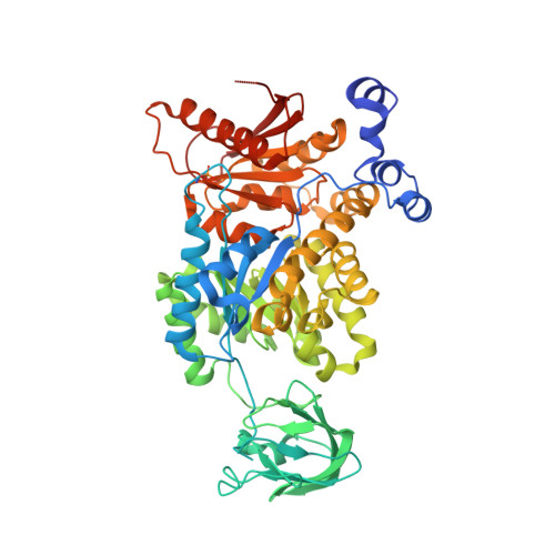
    6ECH, 6ECK

  • PubMed Abstract: 

    Pyruvate kinase is an important enzyme in glycolysis and a key metabolic control point. We recently observed a pyruvate kinase liver isoform (PKL) phosphorylation site at S113 that correlates with insulin resistance in rats on a 3 day high-fat diet (HFD) and suggests additional control points for PKL activity. However, in contrast to the classical model of PKL regulation, neither authentically phosphorylated PKL at S12 nor S113 alone is sufficient to alter enzyme kinetics or structure. Instead, we show that cyclin-dependent kinases (CDKs) are activated by the HFD and responsible for PKL phosphorylation at position S113 in addition to other targets. These CDKs control PKL nuclear retention, alter cytosolic PKL activity, and ultimately influence glucose production. These results change our view of PKL regulation and highlight a previously unrecognized pathway of hepatic CDK activity and metabolic control points that may be important in insulin resistance and type 2 diabetes.


  • Organizational Affiliation

    Department of Cellular and Molecular Physiology, Yale University, New Haven, CT, USA; Department of Systems Biology Institute, Yale University, New Haven, CT, USA.


Macromolecules
Find similar proteins by:  (by identity cutoff)  |  3D Structure
Entity ID: 1
MoleculeChains Sequence LengthOrganismDetailsImage
Pyruvate kinase PKLR
A, B
550Rattus norvegicusMutation(s): 0 
Gene Names: Pklr
EC: 2.7.1.40
UniProt
Find proteins for P12928 (Rattus norvegicus)
Explore P12928 
Go to UniProtKB:  P12928
Entity Groups  
Sequence Clusters30% Identity50% Identity70% Identity90% Identity95% Identity100% Identity
UniProt GroupP12928
Sequence Annotations
Expand
  • Reference Sequence
Small Molecules
Ligands 6 Unique
IDChains Name / Formula / InChI Key2D Diagram3D Interactions
FBP
Query on FBP

Download Ideal Coordinates CCD File 
C [auth A],
J [auth B]
1,6-di-O-phosphono-beta-D-fructofuranose
C6 H14 O12 P2
RNBGYGVWRKECFJ-ARQDHWQXSA-N
FLC
Query on FLC

Download Ideal Coordinates CCD File 
L [auth B]CITRATE ANION
C6 H5 O7
KRKNYBCHXYNGOX-UHFFFAOYSA-K
PGE
Query on PGE

Download Ideal Coordinates CCD File 
N [auth B]TRIETHYLENE GLYCOL
C6 H14 O4
ZIBGPFATKBEMQZ-UHFFFAOYSA-N
PEG
Query on PEG

Download Ideal Coordinates CCD File 
F [auth A],
M [auth B]
DI(HYDROXYETHYL)ETHER
C4 H10 O3
MTHSVFCYNBDYFN-UHFFFAOYSA-N
GOL
Query on GOL

Download Ideal Coordinates CCD File 
D [auth A],
E [auth A],
K [auth B]
GLYCEROL
C3 H8 O3
PEDCQBHIVMGVHV-UHFFFAOYSA-N
EDO
Query on EDO

Download Ideal Coordinates CCD File 
G [auth A],
H [auth A],
I [auth A]
1,2-ETHANEDIOL
C2 H6 O2
LYCAIKOWRPUZTN-UHFFFAOYSA-N
Modified Residues  1 Unique
IDChains TypeFormula2D DiagramParent
SEP
Query on SEP
A, B
L-PEPTIDE LINKINGC3 H8 N O6 PSER
Experimental Data & Validation

Experimental Data

  • Method: X-RAY DIFFRACTION
  • Resolution: 2.36 Å
  • R-Value Free: 0.275 
  • R-Value Work: 0.203 
  • R-Value Observed: 0.207 
  • Space Group: P 21 21 2
Unit Cell:
Length ( Å )Angle ( ˚ )
a = 97.774α = 90
b = 111.843β = 90
c = 127.507γ = 90
Software Package:
Software NamePurpose
HKL-2000data reduction
Aimlessdata scaling
PHASERphasing
REFMACrefinement
PDB_EXTRACTdata extraction

Structure Validation

View Full Validation Report


Ligand Structure Quality Assessment 


Entry History 

Deposition Data

Revision History  (Full details and data files)

  • Version 1.0: 2019-12-04
    Type: Initial release
  • Version 1.1: 2019-12-25
    Changes: Database references
  • Version 1.2: 2020-07-29
    Type: Remediation
    Reason: Carbohydrate remediation
    Changes: Data collection, Derived calculations, Structure summary
  • Version 1.3: 2023-10-11
    Changes: Data collection, Database references, Refinement description, Structure summary