6D66

Crystal structure of the human dual specificity 1 catalytic domain (C258S) as a maltose binding protein fusion in complex with the designed AR protein mbp3_16


Experimental Data Snapshot

  • Method: X-RAY DIFFRACTION
  • Resolution: 2.23 Å
  • R-Value Free: 0.202 
  • R-Value Work: 0.159 
  • R-Value Observed: 0.162 

wwPDB Validation   3D Report Full Report


This is version 1.1 of the entry. See complete history


Literature

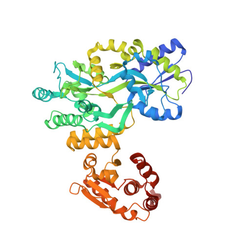
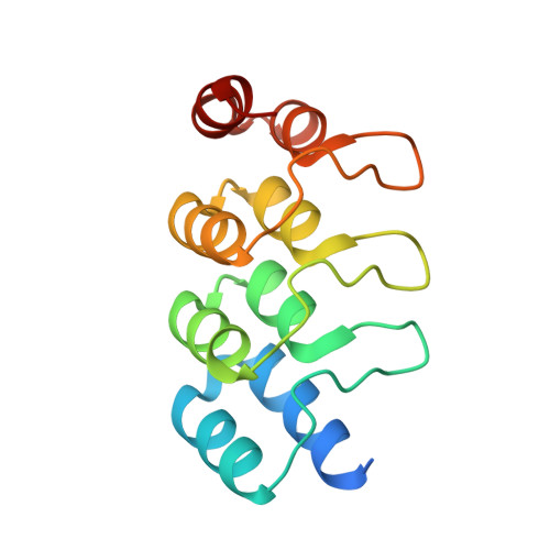
MBP-binding DARPins facilitate the crystallization of an MBP fusion protein.

Gumpena, R.Lountos, G.T.Waugh, D.S.

(2018) Acta Crystallogr F Struct Biol Commun 74: 549-557

  • DOI: https://doi.org/10.1107/S2053230X18009901
  • Primary Citation of Related Structures:  
    6D65, 6D66, 6D67

  • PubMed Abstract: 

    The production of high-quality crystals is the main bottleneck in determining the structures of proteins using X-ray crystallography. In addition to being recognized as a very effective solubility-enhancing fusion partner, Escherichia coli maltose-binding protein (MBP) has also been successfully employed as a `fixed-arm' crystallization chaperone in more than 100 cases. Here, it is reported that designed ankyrin-repeat proteins (DARPins) that bind with high affinity to MBP can promote the crystallization of an MBP fusion protein when the fusion protein alone fails to produce diffraction-quality crystals. As a proof of principle, three different co-crystal structures of MBP fused to the catalytic domain of human dual-specificity phosphatase 1 in complex with DARPins are reported.


  • Organizational Affiliation

    Macromolecular Crystallography Laboratory, Center for Cancer Research, National Cancer Institute at Frederick, Frederick, MD 21702, USA.


Macromolecules
Find similar proteins by:  (by identity cutoff)  |  3D Structure
Entity ID: 1
MoleculeChains Sequence LengthOrganismDetailsImage
Maltose-binding periplasmic protein,Dual specificity protein phosphatase 1520Escherichia coli K-12Homo sapiens
This entity is chimeric
Mutation(s): 9 
Gene Names: malEb4034JW3994DUSP1CL100MKP1PTPN10VH1
EC: 3.1.3.16 (PDB Primary Data), 3.1.3.48 (PDB Primary Data)
UniProt & NIH Common Fund Data Resources
Find proteins for P0AEX9 (Escherichia coli (strain K12))
Explore P0AEX9 
Go to UniProtKB:  P0AEX9
Find proteins for P28562 (Homo sapiens)
Explore P28562 
Go to UniProtKB:  P28562
PHAROS:  P28562
GTEx:  ENSG00000120129 
Entity Groups  
Sequence Clusters30% Identity50% Identity70% Identity90% Identity95% Identity100% Identity
UniProt GroupsP28562P0AEX9
Sequence Annotations
Expand
  • Reference Sequence
Find similar proteins by:  (by identity cutoff)  |  3D Structure
Entity ID: 2
MoleculeChains Sequence LengthOrganismDetailsImage
Designed AR protein mbp3_16136synthetic constructMutation(s): 0 
Entity Groups  
Sequence Clusters30% Identity50% Identity70% Identity90% Identity95% Identity100% Identity
Sequence Annotations
Expand
  • Reference Sequence
Small Molecules
Ligands 7 Unique
IDChains Name / Formula / InChI Key2D Diagram3D Interactions
PG4
Query on PG4

Download Ideal Coordinates CCD File 
J [auth A]TETRAETHYLENE GLYCOL
C8 H18 O5
UWHCKJMYHZGTIT-UHFFFAOYSA-N
PGE
Query on PGE

Download Ideal Coordinates CCD File 
F [auth A],
M [auth A],
Q [auth A]
TRIETHYLENE GLYCOL
C6 H14 O4
ZIBGPFATKBEMQZ-UHFFFAOYSA-N
PEG
Query on PEG

Download Ideal Coordinates CCD File 
C [auth A],
S [auth A],
X [auth B]
DI(HYDROXYETHYL)ETHER
C4 H10 O3
MTHSVFCYNBDYFN-UHFFFAOYSA-N
PO4
Query on PO4

Download Ideal Coordinates CCD File 
D [auth A]PHOSPHATE ION
O4 P
NBIIXXVUZAFLBC-UHFFFAOYSA-K
DAL
Query on DAL

Download Ideal Coordinates CCD File 
Y [auth B]D-ALANINE
C3 H7 N O2
QNAYBMKLOCPYGJ-UWTATZPHSA-N
GLY
Query on GLY

Download Ideal Coordinates CCD File 
E [auth A],
N [auth A],
O [auth A]
GLYCINE
C2 H5 N O2
DHMQDGOQFOQNFH-UHFFFAOYSA-N
EDO
Query on EDO

Download Ideal Coordinates CCD File 
G [auth A]
H [auth A]
I [auth A]
K [auth A]
L [auth A]
G [auth A],
H [auth A],
I [auth A],
K [auth A],
L [auth A],
P [auth A],
R [auth A],
T [auth A],
U [auth A],
V [auth B],
W [auth B],
Z [auth B]
1,2-ETHANEDIOL
C2 H6 O2
LYCAIKOWRPUZTN-UHFFFAOYSA-N
Experimental Data & Validation

Experimental Data

  • Method: X-RAY DIFFRACTION
  • Resolution: 2.23 Å
  • R-Value Free: 0.202 
  • R-Value Work: 0.159 
  • R-Value Observed: 0.162 
  • Space Group: P 41 21 2
Unit Cell:
Length ( Å )Angle ( ˚ )
a = 79.967α = 90
b = 79.967β = 90
c = 265.797γ = 90
Software Package:
Software NamePurpose
PHENIXrefinement
HKL-3000data reduction
HKL-3000data scaling
PHASERphasing

Structure Validation

View Full Validation Report


Entry History 

Deposition Data

Revision History  (Full details and data files)

  • Version 1.0: 2018-09-19
    Type: Initial release
  • Version 1.1: 2023-10-04
    Changes: Data collection, Database references, Refinement description