6ADI

Crystal Structures of IDH2 R140Q in complex with AG-881


Experimental Data Snapshot

  • Method: X-RAY DIFFRACTION
  • Resolution: 1.97 Å
  • R-Value Free: 0.226 
  • R-Value Work: 0.206 
  • R-Value Observed: 0.207 

wwPDB Validation   3D Report Full Report


Ligand Structure Quality Assessment 


This is version 1.2 of the entry. See complete history


Literature

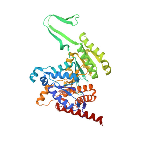
Crystal structures of pan-IDH inhibitor AG-881 in complex with mutant human IDH1 and IDH2

Ma, R.Yun, C.H.

(2018) Biochem Biophys Res Commun 503: 2912-2917

  • DOI: https://doi.org/10.1016/j.bbrc.2018.08.068
  • Primary Citation of Related Structures:  
    6ADG, 6ADI

  • PubMed Abstract: 

    Some mutations of isocitrate dehydrogenase 1 and 2 observed in multiple kinds of malignant tumors can lead to a neomorphic enzyme activity that converts alpha-ketoglutarate (α-KG) to 2-hydroxyglutarate (2-HG). As an oncometabolite, 2-HG can cause epigenetic changes and impair cell differentiation. Inhibiting the activity of isocitrate dehydrogenase mutants (mIDH) is considered to be an effective therapy for the treatment of mIDH positive cancers, including glioma and acute myeloid leukemia (AML). The presently disclosed allosteric inhibitors work only on one of the mIDH1 and mIDH2, and it is shown that mIDH1 and mIDH2 have different allosteric inhibition pockets. However, AG-881 from Agios Pharmaceuticals was found to be a pan-IDH inhibitor against both mIDH1 and mIDH2, and is undergoing Phase I clinical trials for tumors with an IDH1 and/or IDH2 mutation. To understand the binding mode of AG-881 to mIDHs, we solved the crystal structures of IDH1-R132H/NADPH/AG-881 and IDH2-R140Q/NADPH/AG-881 complexes, and acquired the IC 50 values of AG-881 for IDH1-R132H and IDH2-R140Q homodimers after different pre-incubation times. Our data show that AG-881 binds IDH1-R132H and IDH2-R140Q in the same allosteric pockets and that the subtle difference in the pockets of these two proteins may contribute to their remarkably different inhibitory kinetics by AG-881. The structural pharmacological data provided in this report may benefit the future development of pan-IDH inhibitors targeting mIDH1 and mIDH2.


  • Organizational Affiliation

    Department of Biophysics, School of Basic Medical Sciences, Peking University Health Science Center, Beijing 100191, China; Institute of Systems Biomedicine, School of Basic Medical Sciences, Peking University Health Science Center, Beijing 100191, China; Beijing Key Laboratory of Tumor Systems Biology, Peking University Health Science Center, Beijing 100191, China.


Macromolecules
Find similar proteins by:  (by identity cutoff)  |  3D Structure
Entity ID: 1
MoleculeChains Sequence LengthOrganismDetailsImage
Isocitrate dehydrogenase [NADP], mitochondrial
A, B
419Homo sapiensMutation(s): 1 
Gene Names: IDH2
EC: 1.1.1.42
UniProt & NIH Common Fund Data Resources
Find proteins for P48735 (Homo sapiens)
Explore P48735 
Go to UniProtKB:  P48735
PHAROS:  P48735
GTEx:  ENSG00000182054 
Entity Groups  
Sequence Clusters30% Identity50% Identity70% Identity90% Identity95% Identity100% Identity
UniProt GroupP48735
Sequence Annotations
Expand
  • Reference Sequence
Small Molecules
Ligands 2 Unique
IDChains Name / Formula / InChI Key2D Diagram3D Interactions
NDP
Query on NDP

Download Ideal Coordinates CCD File 
C [auth A],
E [auth B]
NADPH DIHYDRO-NICOTINAMIDE-ADENINE-DINUCLEOTIDE PHOSPHATE
C21 H30 N7 O17 P3
ACFIXJIJDZMPPO-NNYOXOHSSA-N
9UO (Subject of Investigation/LOI)
Query on 9UO

Download Ideal Coordinates CCD File 
D [auth A]6-(6-chloropyridin-2-yl)-N2,N4-bis[(2R)-1,1,1-trifluoropropan-2-yl]-1,3,5-triazine-2,4-diamine
C14 H13 Cl F6 N6
QCZAWDGAVJMPTA-RNFRBKRXSA-N
Binding Affinity Annotations 
IDSourceBinding Affinity
9UO BindingDB:  6ADI IC50: min: 7, max: 130 (nM) from 4 assay(s)
Binding MOAD:  6ADI IC50: 31.7 (nM) from 1 assay(s)
Experimental Data & Validation

Experimental Data

  • Method: X-RAY DIFFRACTION
  • Resolution: 1.97 Å
  • R-Value Free: 0.226 
  • R-Value Work: 0.206 
  • R-Value Observed: 0.207 
  • Space Group: P 21 21 21
Unit Cell:
Length ( Å )Angle ( ˚ )
a = 57.781α = 90
b = 118.419β = 90
c = 126.082γ = 90
Software Package:
Software NamePurpose
PHENIXrefinement
DENZOdata reduction
HKL-3000data scaling
PHASERphasing

Structure Validation

View Full Validation Report


Ligand Structure Quality Assessment 


Entry History 

Deposition Data

Revision History  (Full details and data files)

  • Version 1.0: 2018-09-05
    Type: Initial release
  • Version 1.1: 2018-09-26
    Changes: Data collection, Database references
  • Version 1.2: 2023-11-22
    Changes: Data collection, Database references, Refinement description