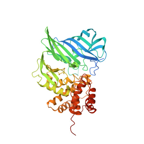
Two-domain aminopeptidase of M1 family: Structural features for substrate binding and gating in absence of C-terminal domain.
Agrawal, R., Goyal, V.D., Kumar, A., Gaur, N.K., Jamdar, S.N., Kumar, A., Makde, R.D.(2019) J Struct Biol 208: 51-60
- PubMed: 31351924
- DOI: https://doi.org/10.1016/j.jsb.2019.07.010
- Primary Citation of Related Structures:
6A8Z, 6IFG - PubMed Abstract:
Zinc metallopeptidases of the M1 family (M1 peptidases) with unique metal binding motif HEXXH(X) 18 E regulate many important biological processes such as tumor growth, angiogenesis, hormone regulation, and immune cell development. Typically, these enzymes exist in three-domain [N-terminal domain (N-domain), catalytic domain, and C-terminal domain (C-domain)] or four-domain (N-domain, catalytic domain, middle domain, and C-domain) format in which N-domain and catalytic domain are more conserved. The C-domain plays important roles in substrate binding and gating. In this study we report the first structure of a two-domain (N-domain and catalytic domain) M1 peptidase at 2.05 Å resolution. Despite the lack of C-domain, the enzyme is active and prefers peptide substrates with large hydrophobic N-terminal residues. Its substrate-bound structure was determined at 1.9 Å resolution. Structural analyses supported by site directed mutagenesis and molecular dynamics simulations reveal structural features that could compensate for the lack of C-domain. A unique loop insertion (loop A) in the N-domain has important roles in gating and desolvation of active site. Three Arg residues of the catalytic domain are involved in substrate-binding roles typically played by positively charged residues of C-domain in other M1 peptidases. Further, its unique exopeptidase sequence motif, LALET, creates a more hydrophobic environment at the S1 subsite (which binds N-terminal residue of the substrate in aminopeptidases) than the more common GXMEN motif in the family. This leads to high affinity for large hydrophobic residues in the S1 subsite, which contributes towards efficient substrate binding in absence of C-domain.
Organizational Affiliation:
Discipline of Biosciences and Biomedical Engineering, Indian Institute of Technology Indore, Indore 453552, India; High Pressure and Synchrotron Radiation Physics Division, Bhabha Atomic Research Centre, Mumbai 400085, India.


