5ZV2

FGFR-1 in complex with ligand lenvatinib


Experimental Data Snapshot

  • Method: X-RAY DIFFRACTION
  • Resolution: 2.86 Å
  • R-Value Free: 0.269 
  • R-Value Work: 0.213 
  • R-Value Observed: 0.215 

wwPDB Validation   3D Report Full Report


Ligand Structure Quality Assessment 


This is version 1.1 of the entry. See complete history


Literature

Lenvatinib inhibits angiogenesis and tumor fibroblast growth factor signaling pathways in human hepatocellular carcinoma models.

Matsuki, M.Hoshi, T.Yamamoto, Y.Ikemori-Kawada, M.Minoshima, Y.Funahashi, Y.Matsui, J.

(2018) Cancer Med 7: 2641-2653

  • DOI: https://doi.org/10.1002/cam4.1517
  • Primary Citation of Related Structures:  
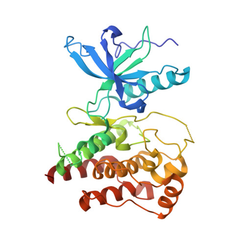
    5ZV2

  • PubMed Abstract: 

    Unresectable hepatocellular carcinoma (uHCC) is one of the most lethal and prevalent cancers worldwide, and current systemic therapeutic options for uHCC are limited. Lenvatinib, a multiple receptor tyrosine kinase inhibitor targeting vascular endothelial growth factor receptors (VEGFRs) and fibroblast growth factor receptors (FGFRs), recently demonstrated a treatment effect on overall survival by statistical confirmation of noninferiority to sorafenib in a phase 3 study of uHCC. Here, we investigated mechanisms underlying the antitumor activity of lenvatinib in preclinical HCC models. In vitro proliferation assay of nine human HCC cell lines showed that lenvatinib selectively inhibited proliferation of FGF signal-activated HCC cells including FGF19-expressing Hep3B2.1-7. Lenvatinib suppressed phosphorylation of FRS2, a substrate of FGFR1-4, in these cells in a concentration-dependent manner. Lenvatinib inhibited in vivo tumor growth in Hep3B2.1-7 and SNU-398 xenografts and decreased phosphorylation of FRS2 and Erk1/2 within the tumor tissues. Lenvatinib also exerted antitumor activity and potently reduced tumor microvessel density in PLC/PRF/5 xenograft model and two HCC patient-derived xenograft models. These results suggest that lenvatinib has antitumor activity consistently across diverse HCC models, and that targeting of tumor FGF signaling pathways and anti-angiogenic activity underlies its antitumor activity against HCC tumors.


  • Organizational Affiliation

    Tsukuba Research Laboratories, Eisai Co., Ltd., Ibaraki, Japan.


Macromolecules
Find similar proteins by:  (by identity cutoff)  |  3D Structure
Entity ID: 1
MoleculeChains Sequence LengthOrganismDetailsImage
Fibroblast growth factor receptor 1
A, B
305Homo sapiensMutation(s): 1 
Gene Names: FGFR1BFGFRCEKFGFBRFLGFLT2HBGFR
EC: 2.7.10.1
UniProt & NIH Common Fund Data Resources
Find proteins for P11362 (Homo sapiens)
Explore P11362 
Go to UniProtKB:  P11362
PHAROS:  P11362
GTEx:  ENSG00000077782 
Entity Groups  
Sequence Clusters30% Identity50% Identity70% Identity90% Identity95% Identity100% Identity
UniProt GroupP11362
Sequence Annotations
Expand
  • Reference Sequence
Small Molecules
Ligands 1 Unique
IDChains Name / Formula / InChI Key2D Diagram3D Interactions
LEV
Query on LEV

Download Ideal Coordinates CCD File 
C [auth A],
D [auth B]
4-{3-chloro-4-[(cyclopropylcarbamoyl)amino]phenoxy}-7-methoxyquinoline-6-carboxamide
C21 H19 Cl N4 O4
WOSKHXYHFSIKNG-UHFFFAOYSA-N
Experimental Data & Validation

Experimental Data

  • Method: X-RAY DIFFRACTION
  • Resolution: 2.86 Å
  • R-Value Free: 0.269 
  • R-Value Work: 0.213 
  • R-Value Observed: 0.215 
  • Space Group: C 1 2 1
Unit Cell:
Length ( Å )Angle ( ˚ )
a = 210.214α = 90
b = 57.464β = 106.75
c = 65.955γ = 90
Software Package:
Software NamePurpose
REFMACrefinement
XDSdata reduction
XSCALEdata scaling
REFMACphasing

Structure Validation

View Full Validation Report


Ligand Structure Quality Assessment 


Entry History 

Deposition Data

Revision History  (Full details and data files)

  • Version 1.0: 2018-07-11
    Type: Initial release
  • Version 1.1: 2023-11-22
    Changes: Advisory, Data collection, Database references, Refinement description