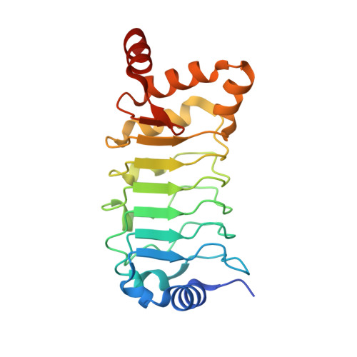
5YXM

Crystal structure of Chlamydomonas Outer Arm Dynein Light Chain 1


Experimental Data Snapshot

  • Method: X-RAY DIFFRACTION
  • Resolution: 1.54 Å
  • R-Value Free: 0.189 
  • R-Value Work: 0.169 
  • R-Value Observed: 0.170 

wwPDB Validation   3D Report Full Report


This is version 1.2 of the entry. See complete history


Literature

Structural atlas of dynein motors at atomic resolution.

Toda, A.Tanaka, H.Kurisu, G.

(2018) Biophys Rev 10: 677-686

  • DOI: https://doi.org/10.1007/s12551-018-0402-y
  • Primary Citation of Related Structures:  
    5YXM

  • PubMed Abstract: 

    Dynein motors are biologically important bio-nanomachines, and many atomic resolution structures of cytoplasmic dynein components from different organisms have been analyzed by X-ray crystallography, cryo-EM, and NMR spectroscopy. This review provides a historical perspective of structural studies of cytoplasmic and axonemal dynein including accessory proteins. We describe representative structural studies of every component of dynein and summarize them as a structural atlas that classifies the cytoplasmic and axonemal dyneins. Based on our review of all dynein structures in the Protein Data Bank, we raise two important points for understanding the two types of dynein motor and discuss the potential prospects of future structural studies.


  • Organizational Affiliation

    Institute for Protein Research, Osaka University, Suita, Osaka, 565-0871, Japan.


Macromolecules
Find similar proteins by:  (by identity cutoff)  |  3D Structure
Entity ID: 1
MoleculeChains Sequence LengthOrganismDetailsImage
Dynein light chain 1, axonemal203Chlamydomonas reinhardtiiMutation(s): 0 
Gene Names: LC1CHLREDRAFT_186669
UniProt
Find proteins for Q9XHH2 (Chlamydomonas reinhardtii)
Explore Q9XHH2 
Go to UniProtKB:  Q9XHH2
Entity Groups  
Sequence Clusters30% Identity50% Identity70% Identity90% Identity95% Identity100% Identity
UniProt GroupQ9XHH2
Sequence Annotations
Expand
  • Reference Sequence
Experimental Data & Validation

Experimental Data

  • Method: X-RAY DIFFRACTION
  • Resolution: 1.54 Å
  • R-Value Free: 0.189 
  • R-Value Work: 0.169 
  • R-Value Observed: 0.170 
  • Space Group: P 21 21 21
Unit Cell:
Length ( Å )Angle ( ˚ )
a = 53.536α = 90
b = 69.23β = 90
c = 74.775γ = 90
Software Package:
Software NamePurpose
PHENIXrefinement
HKL-2000data scaling
BALBESphasing
Cootmodel building
HKL-2000data reduction

Structure Validation

View Full Validation Report


Entry History & Funding Information

Deposition Data


Funding OrganizationLocationGrant Number
Grant-in-Aid for JSPS FellowsJapan17J075530
Grant-in-Aid for Scientific Research(B)Japan26291014

Revision History  (Full details and data files)

  • Version 1.0: 2018-03-14
    Type: Initial release
  • Version 1.1: 2018-04-25
    Changes: Data collection, Database references
  • Version 1.2: 2023-11-22
    Changes: Data collection, Database references, Refinement description