5Y8B

Periplasmic heme-binding protein RhuT from Roseiflexus sp. RS-1 in apo form


Experimental Data Snapshot

  • Method: X-RAY DIFFRACTION
  • Resolution: 2.40 Å
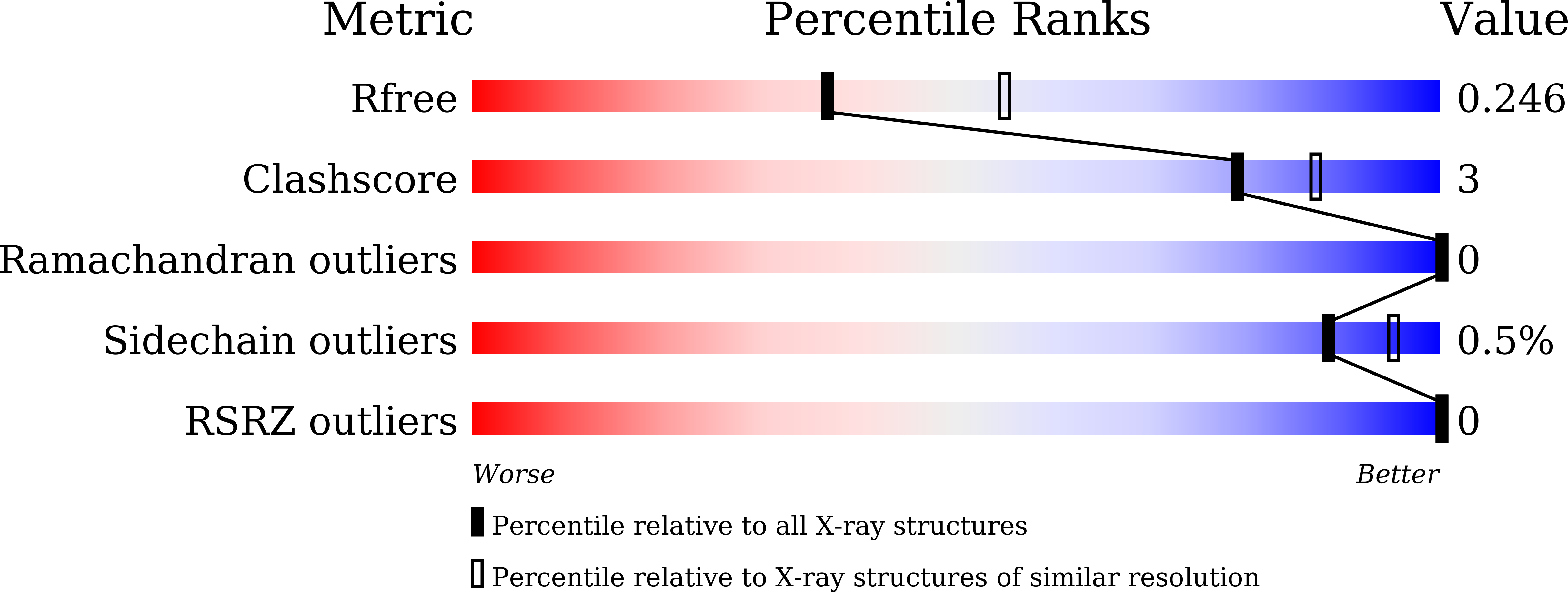
  • R-Value Free: 0.246 
  • R-Value Work: 0.180 
  • R-Value Observed: 0.183 

wwPDB Validation   3D Report Full Report


This is version 1.2 of the entry. See complete history


Literature

Structural basis for binding and transfer of heme in bacterial heme-acquisition systems

Naoe, Y.Nakamura, N.Rahman, M.M.Tosha, T.Nagatoishi, S.Tsumoto, K.Shiro, Y.Sugimoto, H.

(2017) Proteins 85: 2217-2230

  • DOI: https://doi.org/10.1002/prot.25386
  • Primary Citation of Related Structures:  
    5GIZ, 5GJ3, 5Y89, 5Y8A, 5Y8B

  • PubMed Abstract: 

    Periplasmic heme-binding proteins (PBPs) in Gram-negative bacteria are components of the heme acquisition system. These proteins shuttle heme across the periplasmic space from outer membrane receptors to ATP-binding cassette (ABC) heme importers located in the inner-membrane. In the present study, we characterized the structures of PBPs found in the pathogen Burkholderia cenocepacia (BhuT) and in the thermophile Roseiflexus sp. RS-1 (RhuT) in the heme-free and heme-bound forms. The conserved motif, in which a well-conserved Tyr interacts with the nearby Arg coordinates on heme iron, was observed in both PBPs. The heme was recognized by its surroundings in a variety of manners including hydrophobic interactions and hydrogen bonds, which was confirmed by isothermal titration calorimetry. Furthermore, this study of 3 forms of BhuT allowed the first structural comparison and showed that the heme-binding cleft of BhuT adopts an "open" state in the heme-free and 2-heme-bound forms, and a "closed" state in the one-heme-bound form with unique conformational changes. Such a conformational change might adjust the interaction of the heme(s) with the residues in PBP and facilitate the transfer of the heme into the translocation channel of the importer.


  • Organizational Affiliation

    Biometal Science Laboratory, RIKEN SPring-8 Center, 1-1-1 Kouto, Sayo, Hyogo, 679-5148, Japan.


Macromolecules
Find similar proteins by:  (by identity cutoff)  |  3D Structure
Entity ID: 1
MoleculeChains Sequence LengthOrganismDetailsImage
Periplasmic binding protein270Roseiflexus sp. RS-1Mutation(s): 0 
Gene Names: RoseRS_3565
UniProt
Find proteins for A5UZ69 (Roseiflexus sp. (strain RS-1))
Explore A5UZ69 
Go to UniProtKB:  A5UZ69
Entity Groups  
Sequence Clusters30% Identity50% Identity70% Identity90% Identity95% Identity100% Identity
UniProt GroupA5UZ69
Sequence Annotations
Expand
  • Reference Sequence
Experimental Data & Validation

Experimental Data

  • Method: X-RAY DIFFRACTION
  • Resolution: 2.40 Å
  • R-Value Free: 0.246 
  • R-Value Work: 0.180 
  • R-Value Observed: 0.183 
  • Space Group: P 31 2 1
Unit Cell:
Length ( Å )Angle ( ˚ )
a = 71.039α = 90
b = 71.039β = 90
c = 78.206γ = 120
Software Package:
Software NamePurpose
SCALEPACKdata scaling
REFMACrefinement
PDB_EXTRACTdata extraction
MOLREPphasing
HKL-2000data reduction

Structure Validation

View Full Validation Report


Entry History & Funding Information

Deposition Data


Funding OrganizationLocationGrant Number
Ministry of Education, Culture, Sports, Science and Technology (Japan)JapanJP23121531
Ministry of Education, Culture, Sports, Science and Technology (Japan)JapanJP25121739
Ministry of Education, Culture, Sports, Science and Technology (Japan)JapanJP15H01655

Revision History  (Full details and data files)

  • Version 1.0: 2017-10-11
    Type: Initial release
  • Version 1.1: 2017-11-22
    Changes: Database references
  • Version 1.2: 2023-11-22
    Changes: Data collection, Database references, Derived calculations, Refinement description