5WQW

X-ray structure of catalytic domain of autolysin from Clostridium perfringens


Experimental Data Snapshot

  • Method: X-RAY DIFFRACTION
  • Resolution: 1.76 Å
  • R-Value Free: 0.192 
  • R-Value Work: 0.161 
  • R-Value Observed: 0.162 

wwPDB Validation   3D Report Full Report


This is version 1.2 of the entry. See complete history


Literature

Structural and biochemical characterization of the Clostridium perfringens autolysin catalytic domain

Tamai, E.Sekiya, H.Goda, E.Makihata, N.Maki, J.Yoshida, H.Kamitori, S.

(2017) FEBS Lett 591: 231-239

  • DOI: https://doi.org/10.1002/1873-3468.12515
  • Primary Citation of Related Structures:  
    5WQW

  • PubMed Abstract: 

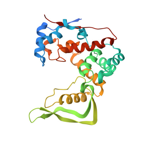
    Bacterial autolysins can partially hydrolyze cell wall peptidoglycans into small sections to regulate cell separation/division and the growth phase. Clostridium perfringens autolysin (Acp) has an N-terminal cell wall-binding domain and a C-terminal catalytic domain with glucosaminidase activity that belongs to the glycoside hydrolase 73 family. Here, we determined the X-ray structure of the Acp catalytic domain (AcpCD) at 1.76 Å resolution. AcpCD has a unique crescent-shaped structure, forming a deep groove for substrate-binding at the center of the protein. The modeling study of the enzyme/substrate complex demonstrated that the length of the substrate-binding groove is closely related to the glucosaminidase activity. Mutagenesis analysis showed that AcpCD likely adopts a neighboring-group mechanism for the catalytic reaction.


  • Organizational Affiliation

    Department of Infectious Disease, College of Pharmaceutical Science, Matsuyama University, Bunkyo-cho, Ehime, Japan.


Macromolecules
Find similar proteins by:  (by identity cutoff)  |  3D Structure
Entity ID: 1
MoleculeChains Sequence LengthOrganismDetailsImage
N-acetylglucosaminidase271Clostridium perfringens str. 13Mutation(s): 0 
Gene Names: CPE1231
UniProt
Find proteins for Q8XL11 (Clostridium perfringens (strain 13 / Type A))
Explore Q8XL11 
Go to UniProtKB:  Q8XL11
Entity Groups  
Sequence Clusters30% Identity50% Identity70% Identity90% Identity95% Identity100% Identity
UniProt GroupQ8XL11
Sequence Annotations
Expand
  • Reference Sequence
Experimental Data & Validation

Experimental Data

  • Method: X-RAY DIFFRACTION
  • Resolution: 1.76 Å
  • R-Value Free: 0.192 
  • R-Value Work: 0.161 
  • R-Value Observed: 0.162 
  • Space Group: P 1 21 1
Unit Cell:
Length ( Å )Angle ( ˚ )
a = 50.32α = 90
b = 37.17β = 100.34
c = 64γ = 90
Software Package:
Software NamePurpose
REFMACrefinement
XDSdata reduction
XDSdata scaling
PHENIXphasing

Structure Validation

View Full Validation Report


Entry History & Funding Information

Deposition Data


Funding OrganizationLocationGrant Number
JSPS KAKENHIJapanJP15K06973, JP15K08482

Revision History  (Full details and data files)

  • Version 1.0: 2016-12-07
    Type: Initial release
  • Version 1.1: 2017-02-01
    Changes: Database references
  • Version 1.2: 2024-03-20
    Changes: Data collection, Database references