5WPQ

Cryo-EM structure of mammalian endolysosomal TRPML1 channel in nanodiscs in closed I conformation at 3.64 Angstrom resolution

  • Classification: MEMBRANE PROTEIN
  • Organism(s): Mus musculus
  • Expression System: Homo sapiens
  • Mutation(s): No 
  • Membrane Protein: Yes  OPMPDBTMMemProtMDmpstruc

  • Deposited: 2017-08-07 Released: 2017-10-18 
  • Deposition Author(s): Chen, Q., She, J., Guo, J., Bai, X., Jiang, Y.
  • Funding Organization(s): National Institutes of Health/National Center for Research Resources (NIH/NCRR), Howard Hughes Medical Institute (HHMI), Cancer Prevention and Research Institute of Texas (CPRIT), Murchison Linthicum Scholar in Medical Research fund, Welch Foundation

Experimental Data Snapshot

  • Method: ELECTRON MICROSCOPY
  • Resolution: 3.64 Å
  • Aggregation State: PARTICLE 
  • Reconstruction Method: SINGLE PARTICLE 

wwPDB Validation   3D Report Full Report


This is version 2.1 of the entry. See complete history


Literature

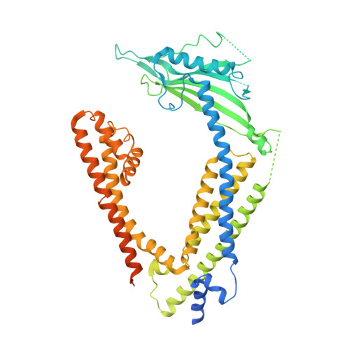
Structure of mammalian endolysosomal TRPML1 channel in nanodiscs.

Chen, Q.She, J.Zeng, W.Guo, J.Xu, H.Bai, X.C.Jiang, Y.

(2017) Nature 550: 415-418

  • DOI: https://doi.org/10.1038/nature24035
  • Primary Citation of Related Structures:  
    5WPQ, 5WPT, 5WPV

  • PubMed Abstract: 

    Transient receptor potential mucolipin 1 (TRPML1) is a cation channel located within endosomal and lysosomal membranes. Ubiquitously expressed in mammalian cells, its loss-of-function mutations are the direct cause of type IV mucolipidosis, an autosomal recessive lysosomal storage disease. Here we present the single-particle electron cryo-microscopy structure of the mouse TRPML1 channel embedded in nanodiscs. Combined with mutagenesis analysis, the TRPML1 structure reveals that phosphatidylinositol-3,5-bisphosphate (PtdIns(3,5)P 2 ) binds to the N terminus of the channel-distal from the pore-and the helix-turn-helix extension between segments S2 and S3 probably couples ligand binding to pore opening. The tightly packed selectivity filter contains multiple ion-binding sites, and the conserved acidic residues form the luminal Ca 2+ -blocking site that confers luminal pH and Ca 2+ modulation on channel conductance. A luminal linker domain forms a fenestrated canopy atop the channel, providing several luminal ion passages to the pore and creating a negative electrostatic trap, with a preference for divalent cations, at the luminal entrance. The structure also reveals two equally distributed S4-S5 linker conformations in the closed channel, suggesting an S4-S5 linker-mediated PtdInsP 2 gating mechanism among TRPML channels.


  • Organizational Affiliation

    Department of Physiology, University of Texas Southwestern Medical Center, Dallas, Texas 75390-9040, USA.


Macromolecules
Find similar proteins by:  (by identity cutoff)  |  3D Structure
Entity ID: 1
MoleculeChains Sequence LengthOrganismDetailsImage
Mucolipin-1
A, B, C, D
592Mus musculusMutation(s): 0 
Gene Names: Mcoln1
Membrane Entity: Yes 
UniProt
Find proteins for Q99J21 (Mus musculus)
Explore Q99J21 
Go to UniProtKB:  Q99J21
Entity Groups  
Sequence Clusters30% Identity50% Identity70% Identity90% Identity95% Identity100% Identity
UniProt GroupQ99J21
Sequence Annotations
Expand
  • Reference Sequence
Oligosaccharides

Help

Entity ID: 2
MoleculeChains Length2D Diagram Glycosylation3D Interactions
2-acetamido-2-deoxy-beta-D-glucopyranose-(1-4)-2-acetamido-2-deoxy-beta-D-glucopyranose
E, F, G, H
2N-Glycosylation
Glycosylation Resources
GlyTouCan:  G42666HT
GlyCosmos:  G42666HT
GlyGen:  G42666HT
Small Molecules
Ligands 1 Unique
IDChains Name / Formula / InChI Key2D Diagram3D Interactions
NA
Query on NA

Download Ideal Coordinates CCD File 
I [auth A]SODIUM ION
Na
FKNQFGJONOIPTF-UHFFFAOYSA-N
Experimental Data & Validation

Experimental Data

  • Method: ELECTRON MICROSCOPY
  • Resolution: 3.64 Å
  • Aggregation State: PARTICLE 
  • Reconstruction Method: SINGLE PARTICLE 
EM Software:
TaskSoftware PackageVersion
RECONSTRUCTIONRELION2

Structure Validation

View Full Validation Report


Entry History & Funding Information

Deposition Data


Funding OrganizationLocationGrant Number
National Institutes of Health/National Center for Research Resources (NIH/NCRR)United StatesGM079179
National Institutes of Health/National Center for Research Resources (NIH/NCRR)United StatesNS062792
National Institutes of Health/National Center for Research Resources (NIH/NCRR)United StatesAR060837
Howard Hughes Medical Institute (HHMI)United States--
Cancer Prevention and Research Institute of Texas (CPRIT)United States--
Murchison Linthicum Scholar in Medical Research fundUnited States--
Welch FoundationUnited StatesI-1578

Revision History  (Full details and data files)

  • Version 1.0: 2017-10-18
    Type: Initial release
  • Version 1.1: 2017-10-25
    Changes: Database references
  • Version 1.2: 2019-04-24
    Changes: Author supporting evidence, Data collection
  • Version 1.3: 2019-11-20
    Changes: Author supporting evidence
  • Version 2.0: 2020-07-29
    Type: Remediation
    Reason: Carbohydrate remediation
    Changes: Atomic model, Data collection, Derived calculations, Structure summary
  • Version 2.1: 2023-04-05
    Changes: Database references, Structure summary