5VC1

Crystal structure of L-selectin lectin/EGF domains


Experimental Data Snapshot

  • Method: X-RAY DIFFRACTION
  • Resolution: 1.94 Å
  • R-Value Free: 0.231 
  • R-Value Work: 0.202 
  • R-Value Observed: 0.203 

wwPDB Validation   3D Report Full Report


This is version 2.1 of the entry. See complete history


Literature

Reducing Macro- and Microheterogeneity of N-Glycans Enables the Crystal Structure of the Lectin and EGF-Like Domains of Human L-Selectin To Be Solved at 1.9 angstrom Resolution.

Wedepohl, S.Dernedde, J.Vahedi-Faridi, A.Tauber, R.Saenger, W.Bulut, H.

(2017) Chembiochem 18: 1338-1345

  • DOI: https://doi.org/10.1002/cbic.201700220
  • Primary Citation of Related Structures:  
    5VC1

  • PubMed Abstract: 

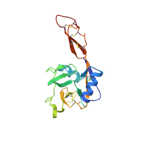
    L-Selectin, a cell-adhesion receptor on the surface of most leukocytes, contains seven N-glycosylation sites. In order to obtain the crystal structure of human L-selectin, we expressed a shortened version of L-selectin comprising the C-type lectin and EGF-like domains (termed LE) and systematically analysed mutations of the three glycosylation sites (Asn22, Asn66 and Asn139) in order to reduce macroheterogeneity. After we further removed microheterogeneity, we obtained crystals that diffracted X-rays up to 1.9 Å from a variant (LE010) with exchanges N22Q and N139Q and one GlcNAc 2 Man 5 N-glycan chain attached to Asn66. Crystal-structure analysis showed that the terminal mannose of GlcNAc 2 Man 5 of one LE010 molecule was coordinated to Ca 2+ in the binding site of a symmetry-related LE010. The orientation of the lectin and EGF-like domain was similar to the described "bent" conformation of E- and P-selectins. The Ca 2+ -binding site reflects the binding mode seen in E- and P-selectin structures co-crystallised with ligands.


  • Organizational Affiliation

    Institut für Laboratoriumsmedizin, Klinische Chemie und Pathobiochemie, Charité Universitätsmedizin Berlin, CVK, Augustenburger Platz 1, 13353, Berlin, Germany.


Macromolecules
Find similar proteins by:  (by identity cutoff)  |  3D Structure
Entity ID: 1
MoleculeChains Sequence LengthOrganismDetailsImage
L-selectin157Homo sapiensMutation(s): 2 
Gene Names: SELLLNHRLYAM1
UniProt & NIH Common Fund Data Resources
Find proteins for P14151 (Homo sapiens)
Explore P14151 
Go to UniProtKB:  P14151
PHAROS:  P14151
GTEx:  ENSG00000188404 
Entity Groups  
Sequence Clusters30% Identity50% Identity70% Identity90% Identity95% Identity100% Identity
UniProt GroupP14151
Sequence Annotations
Expand
  • Reference Sequence
Oligosaccharides

Help

Entity ID: 2
MoleculeChains Length2D Diagram Glycosylation3D Interactions
beta-D-mannopyranose-(1-3)-[beta-D-mannopyranose-(1-6)]alpha-D-mannopyranose-(1-4)-2-acetamido-2-deoxy-beta-D-glucopyranose-(1-4)-2-acetamido-2-deoxy-beta-D-glucopyranose
B
5N-Glycosylation
Glycosylation Resources
GlyTouCan:  G23553FE
GlyCosmos:  G23553FE
GlyGen:  G23553FE
Experimental Data & Validation

Experimental Data

  • Method: X-RAY DIFFRACTION
  • Resolution: 1.94 Å
  • R-Value Free: 0.231 
  • R-Value Work: 0.202 
  • R-Value Observed: 0.203 
  • Space Group: I 2 3
Unit Cell:
Length ( Å )Angle ( ˚ )
a = 118.598α = 90
b = 118.598β = 90
c = 118.598γ = 90
Software Package:
Software NamePurpose
PHENIXrefinement
Cootmodel building
XDSdata scaling
PHENIXphasing
XDSdata reduction

Structure Validation

View Full Validation Report


Entry History & Funding Information

Deposition Data


Funding OrganizationLocationGrant Number
German Research FoundationGermanySFB-449

Revision History  (Full details and data files)

  • Version 1.0: 2017-06-14
    Type: Initial release
  • Version 1.1: 2017-07-12
    Changes: Database references
  • Version 1.2: 2019-10-16
    Changes: Author supporting evidence, Data collection
  • Version 2.0: 2020-07-29
    Type: Remediation
    Reason: Carbohydrate remediation
    Changes: Advisory, Atomic model, Data collection, Derived calculations, Structure summary
  • Version 2.1: 2024-01-17
    Changes: Data collection, Database references, Derived calculations, Refinement description, Structure summary