5UOH

Crystal Structure of Hip1 (Rv2224c) T466A mutant


Experimental Data Snapshot

  • Method: X-RAY DIFFRACTION
  • Resolution: 2.61 Å
  • R-Value Free: 0.258 
  • R-Value Work: 0.217 
  • R-Value Observed: 0.221 

wwPDB Validation   3D Report Full Report


This is version 1.4 of the entry. See complete history


Literature

Structure Determination of Mycobacterium tuberculosis Serine Protease Hip1 (Rv2224c).

Naffin-Olivos, J.L.Daab, A.White, A.Goldfarb, N.E.Milne, A.C.Liu, D.Baikovitz, J.Dunn, B.M.Rengarajan, J.Petsko, G.A.Ringe, D.

(2017) Biochemistry 56: 2304-2314

  • DOI: https://doi.org/10.1021/acs.biochem.6b01066
  • Primary Citation of Related Structures:  
    5UGQ, 5UNO, 5UOH

  • PubMed Abstract: 

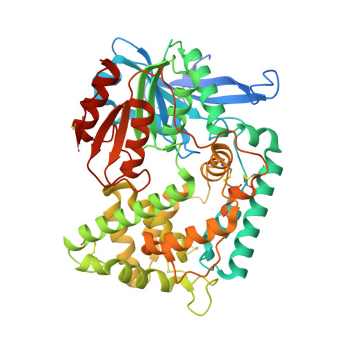
    The Mycobacterium tuberculosis (Mtb) serine protease Hip1 (hydrolase important for pathogenesis; Rv2224c) promotes tuberculosis (TB) pathogenesis by impairing host immune responses through proteolysis of a protein substrate, Mtb GroEL2. The cell surface localization of Hip1 and its immunomodulatory functions make Hip1 a good drug target for new adjunctive immune therapies for TB. Here, we report the crystal structure of Hip1 to a resolution of 2.6 Å and the kinetic studies of the enzyme against model substrates and the protein GroEL2. The structure shows a two-domain protein, one of which contains the catalytic residues that are the signature of a serine protease. Surprisingly, a threonine is located within the active site close enough to hydrogen bond with the catalytic residues Asp463 and His490. Mutation of this residue, Thr466, to alanine established its importance for function. Our studies provide insights into the structure of a member of a novel family of proteases. Knowledge of the Hip1 structure will aid in designing inhibitors that could block Hip1 activity.


  • Organizational Affiliation

    Rosenstiel Basic Medical Sciences Research Center, Brandeis University , Waltham, Massachusetts 02454, United States.


Macromolecules
Find similar proteins by:  (by identity cutoff)  |  3D Structure
Entity ID: 1
MoleculeChains Sequence LengthOrganismDetailsImage
Carboxylesterase A492Mycobacterium tuberculosis H37RvMutation(s): 1 
Gene Names: caeARv2224cMTCY427.05c
EC: 3.1.1
UniProt
Find proteins for P9WHR3 (Mycobacterium tuberculosis (strain ATCC 25618 / H37Rv))
Explore P9WHR3 
Go to UniProtKB:  P9WHR3
Entity Groups  
Sequence Clusters30% Identity50% Identity70% Identity90% Identity95% Identity100% Identity
UniProt GroupP9WHR3
Sequence Annotations
Expand
  • Reference Sequence
Experimental Data & Validation

Experimental Data

  • Method: X-RAY DIFFRACTION
  • Resolution: 2.61 Å
  • R-Value Free: 0.258 
  • R-Value Work: 0.217 
  • R-Value Observed: 0.221 
  • Space Group: P 31 2 1
Unit Cell:
Length ( Å )Angle ( ˚ )
a = 104.249α = 90
b = 104.249β = 90
c = 127.707γ = 120
Software Package:
Software NamePurpose
HKL-2000data scaling
PHENIXrefinement
PDB_EXTRACTdata extraction
SCALEPACKdata scaling
HKL-2000data reduction
PHENIXphasing
DENZOdata reduction

Structure Validation

View Full Validation Report


Entry History & Funding Information

Deposition Data


Funding OrganizationLocationGrant Number
National Institutes of Health/John E. Fogarty International Center (NIH/FIC)United StatesR00TW008043
National Institutes of Health/National Institute Of Allergy and Infectious Diseases (NIH/NIAID)United States5R01AI083366
National Institutes of Health/National Institute of General Medical Sciences (NIH/NIGMS)United StatesGM 32415
National Institutes of Health/National Institute Of Allergy and Infectious Diseases (NIH/NIAID)United StatesR37AI28571

Revision History  (Full details and data files)

  • Version 1.0: 2017-04-12
    Type: Initial release
  • Version 1.1: 2017-05-10
    Changes: Database references
  • Version 1.2: 2017-09-27
    Changes: Author supporting evidence, Refinement description
  • Version 1.3: 2019-12-04
    Changes: Author supporting evidence
  • Version 1.4: 2019-12-11
    Changes: Author supporting evidence