5M30

Structure of TssK from T6SS EAEC in complex with nanobody nb18


Experimental Data Snapshot

  • Method: X-RAY DIFFRACTION
  • Resolution: 2.60 Å
  • R-Value Free: 0.226 
  • R-Value Work: 0.207 
  • R-Value Observed: 0.208 

wwPDB Validation   3D Report Full Report


This is version 1.3 of the entry. See complete history


Literature

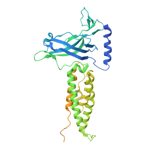
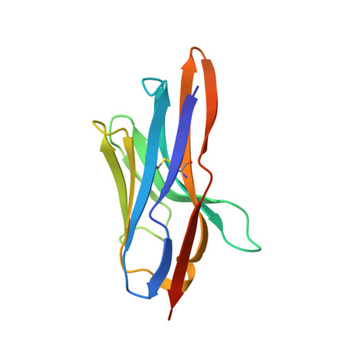
Type VI secretion TssK baseplate protein exhibits structural similarity with phage receptor-binding proteins and evolved to bind the membrane complex.

Nguyen, V.S.Logger, L.Spinelli, S.Legrand, P.Huyen Pham, T.T.Nhung Trinh, T.T.Cherrak, Y.Zoued, A.Desmyter, A.Durand, E.Roussel, A.Kellenberger, C.Cascales, E.Cambillau, C.

(2017) Nat Microbiol 2: 17103-17103

  • DOI: https://doi.org/10.1038/nmicrobiol.2017.103
  • Primary Citation of Related Structures:  
    5M2W, 5M2Y, 5M30, 5MWN

  • PubMed Abstract: 

    The type VI secretion system (T6SS) is a multiprotein machine widespread in Gram-negative bacteria that delivers toxins into both eukaryotic and prokaryotic cells. The mechanism of action of the T6SS is comparable to that of contractile myophages. The T6SS builds a tail-like structure made of an inner tube wrapped by a sheath, assembled under an extended conformation. Contraction of the sheath propels the inner tube towards the target cell. The T6SS tail is assembled on a platform-the baseplate-which is functionally similar to bacteriophage baseplates. In addition, the baseplate docks the tail to a trans-envelope membrane complex that orients the tail towards the target. Here, we report the crystal structure of TssK, a central component of the T6SS baseplate. We show that TssK is composed of three domains, and establish the contribution of each domain to the interaction with TssK partners. Importantly, this study reveals that the N-terminal domain of TssK is structurally homologous to the shoulder domain of phage receptor-binding proteins, and the C-terminal domain binds the membrane complex. We propose that TssK has conserved the domain of attachment to the virion particle but has evolved the reception domain to use the T6SS membrane complex as receptor.


  • Organizational Affiliation

    Architecture et Fonction des Macromolécules Biologiques, Aix-Marseille Université, Campus de Luminy, Case 932, 13288 Marseille Cedex 09, France.


Macromolecules
Find similar proteins by:  (by identity cutoff)  |  3D Structure
Entity ID: 1
MoleculeChains Sequence LengthOrganismDetailsImage
Type VI secretion protein
A, B, C
445Escherichia coliMutation(s): 0 
Gene Names: ACU81_04625
UniProt
Find proteins for D3GU39 (Escherichia coli O44:H18 (strain 042 / EAEC))
Explore D3GU39 
Go to UniProtKB:  D3GU39
Entity Groups  
Sequence Clusters30% Identity50% Identity70% Identity90% Identity95% Identity100% Identity
UniProt GroupD3GU39
Sequence Annotations
Expand
  • Reference Sequence
Find similar proteins by:  (by identity cutoff)  |  3D Structure
Entity ID: 2
MoleculeChains Sequence LengthOrganismDetailsImage
Anti-vesicular stomatitis virus N VHH
D, E, F
125Vicugna pacosMutation(s): 0 
Entity Groups  
Sequence Clusters30% Identity50% Identity70% Identity90% Identity95% Identity100% Identity
Sequence Annotations
Expand
  • Reference Sequence
Experimental Data & Validation

Experimental Data

  • Method: X-RAY DIFFRACTION
  • Resolution: 2.60 Å
  • R-Value Free: 0.226 
  • R-Value Work: 0.207 
  • R-Value Observed: 0.208 
  • Space Group: P 21 21 21
Unit Cell:
Length ( Å )Angle ( ˚ )
a = 93.24α = 90
b = 153.67β = 90
c = 154.81γ = 90
Software Package:
Software NamePurpose
BUSTERrefinement
XDSdata reduction
XSCALEdata scaling
MOLREPphasing

Structure Validation

View Full Validation Report


Entry History & Funding Information

Deposition Data


Funding OrganizationLocationGrant Number
French National Research AgencyFranceANR- 14-CE14-0006-01

Revision History  (Full details and data files)

  • Version 1.0: 2017-06-21
    Type: Initial release
  • Version 1.1: 2017-06-28
    Changes: Database references
  • Version 1.2: 2017-07-05
    Changes: Database references
  • Version 1.3: 2017-09-06
    Changes: Author supporting evidence