5KER

Deer mouse recombinant hemoglobin from high altitude species


Experimental Data Snapshot

  • Method: X-RAY DIFFRACTION
  • Resolution: 2.20 Å
  • R-Value Free: 0.202 
  • R-Value Work: 0.193 
  • R-Value Observed: 0.194 

wwPDB Validation   3D Report Full Report


Ligand Structure Quality Assessment 


This is version 1.3 of the entry. See complete history


Literature

Alteration of the alpha 1 beta 2/ alpha 2 beta 1 subunit interface contributes to the increased hemoglobin-oxygen affinity of high-altitude deer mice.

Inoguchi, N.Mizuno, N.Baba, S.Kumasaka, T.Natarajan, C.Storz, J.F.Moriyama, H.

(2017) PLoS One 12: e0174921-e0174921

  • DOI: https://doi.org/10.1371/journal.pone.0174921
  • Primary Citation of Related Structures:  
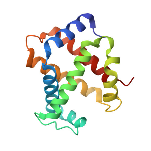
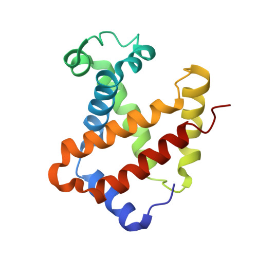
    5KER

  • PubMed Abstract: 

    Deer mice (Peromyscus maniculatus) that are native to high altitudes in the Rocky Mountains have evolved hemoglobins with an increased oxygen-binding affinity relative to those of lowland conspecifics. To elucidate the molecular mechanisms responsible for the evolved increase in hemoglobin-oxygen affinity, the crystal structure of the highland hemoglobin variant was solved and compared with the previously reported structure for the lowland variant. Highland hemoglobin yielded at least two crystal types, in which the longest axes were 507 and 230 Å. Using the smaller unit cell crystal, the structure was solved at 2.2 Å resolution. The asymmetric unit contained two tetrameric hemoglobin molecules. The analyses revealed that αPro50 in the highland hemoglobin variant promoted a stable interaction between αHis45 and heme that was not seen in the αHis50 lowland variant. The αPro50 mutation also altered the nature of atomic contacts at the α1β2/α2β1 intersubunit interfaces. These results demonstrate how affinity-altering changes in intersubunit interactions can be produced by mutations at structurally remote sites.


  • Organizational Affiliation

    School of Biological Sciences, University of Nebraska, Lincoln, Nebraska, United States of America.


Macromolecules
Find similar proteins by:  (by identity cutoff)  |  3D Structure
Entity ID: 1
MoleculeChains Sequence LengthOrganismDetailsImage
Alpha-globinA [auth C],
C [auth E],
E [auth G],
G [auth A]
141Peromyscus maniculatusMutation(s): 0 
Gene Names: HBAHbaHBAT1
UniProt
Find proteins for A4ZQ87 (Peromyscus maniculatus)
Explore A4ZQ87 
Go to UniProtKB:  A4ZQ87
Entity Groups  
Sequence Clusters30% Identity50% Identity70% Identity90% Identity95% Identity100% Identity
UniProt GroupA4ZQ87
Sequence Annotations
Expand
  • Reference Sequence
Find similar proteins by:  (by identity cutoff)  |  3D Structure
Entity ID: 2
MoleculeChains Sequence LengthOrganismDetailsImage
Beta globinB [auth D],
D [auth F],
F [auth H],
H [auth B]
146Peromyscus maniculatusMutation(s): 0 
Gene Names: HBBHBB-T1HBB-T2
UniProt
Find proteins for B7SBL4 (Peromyscus maniculatus)
Explore B7SBL4 
Go to UniProtKB:  B7SBL4
Entity Groups  
Sequence Clusters30% Identity50% Identity70% Identity90% Identity95% Identity100% Identity
UniProt GroupB7SBL4
Sequence Annotations
Expand
  • Reference Sequence
Experimental Data & Validation

Experimental Data

  • Method: X-RAY DIFFRACTION
  • Resolution: 2.20 Å
  • R-Value Free: 0.202 
  • R-Value Work: 0.193 
  • R-Value Observed: 0.194 
  • Space Group: P 1 21 1
Unit Cell:
Length ( Å )Angle ( ˚ )
a = 53.182α = 90
b = 229.618β = 119.12
c = 53.277γ = 90
Software Package:
Software NamePurpose
PHENIXrefinement
HKL-2000data reduction
HKL-2000data scaling
PHENIXphasing

Structure Validation

View Full Validation Report


Ligand Structure Quality Assessment 


Entry History 

Deposition Data

Revision History  (Full details and data files)

  • Version 1.0: 2017-04-05
    Type: Initial release
  • Version 1.1: 2017-04-12
    Changes: Database references
  • Version 1.2: 2017-04-19
    Changes: Derived calculations
  • Version 1.3: 2023-09-27
    Changes: Data collection, Database references, Derived calculations, Refinement description