5JDA

Bacillus cereus CotH kinase plus Mg2+/AMP


Experimental Data Snapshot

  • Method: X-RAY DIFFRACTION
  • Resolution: 1.40 Å
  • R-Value Free: 0.184 
  • R-Value Work: 0.170 
  • R-Value Observed: 0.171 

wwPDB Validation   3D Report Full Report


Ligand Structure Quality Assessment 


This is version 1.3 of the entry. See complete history


Literature

Phosphorylation of spore coat proteins by a family of atypical protein kinases.

Nguyen, K.B.Sreelatha, A.Durrant, E.S.Lopez-Garrido, J.Muszewska, A.Dudkiewicz, M.Grynberg, M.Yee, S.Pogliano, K.Tomchick, D.R.Pawowski, K.Dixon, J.E.Tagliabracci, V.S.

(2016) Proc Natl Acad Sci U S A 113: E3482-E3491

  • DOI: https://doi.org/10.1073/pnas.1605917113
  • Primary Citation of Related Structures:  
    5JD9, 5JDA

  • PubMed Abstract: 

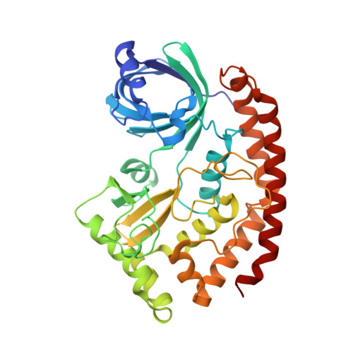
    The modification of proteins by phosphorylation occurs in all life forms and is catalyzed by a large superfamily of enzymes known as protein kinases. We recently discovered a family of secretory pathway kinases that phosphorylate extracellular proteins. One member, family with sequence similarity 20C (Fam20C), is the physiological Golgi casein kinase. While examining distantly related protein sequences, we observed low levels of identity between the spore coat protein H (CotH), and the Fam20C-related secretory pathway kinases. CotH is a component of the spore in many bacterial and eukaryotic species, and is required for efficient germination of spores in Bacillus subtilis; however, the mechanism by which CotH affects germination is unclear. Here, we show that CotH is a protein kinase. The crystal structure of CotH reveals an atypical protein kinase-like fold with a unique mode of ATP binding. Examination of the genes neighboring cotH in B. subtilis led us to identify two spore coat proteins, CotB and CotG, as CotH substrates. Furthermore, we show that CotH-dependent phosphorylation of CotB and CotG is required for the efficient germination of B. subtilis spores. Collectively, our results define a family of atypical protein kinases and reveal an unexpected role for protein phosphorylation in spore biology.


  • Organizational Affiliation

    Department of Pharmacology, University of California, San Diego, La Jolla, CA 92093;


Macromolecules
Find similar proteins by:  (by identity cutoff)  |  3D Structure
Entity ID: 1
MoleculeChains Sequence LengthOrganismDetailsImage
Spore coat protein H359Bacillus cereus ATCC 10987Mutation(s): 0 
Gene Names: cotHBCE_2115
EC: 2.7.11.1
UniProt
Find proteins for Q739M5 (Bacillus cereus (strain ATCC 10987 / NRS 248))
Explore Q739M5 
Go to UniProtKB:  Q739M5
Entity Groups  
Sequence Clusters30% Identity50% Identity70% Identity90% Identity95% Identity100% Identity
UniProt GroupQ739M5
Sequence Annotations
Expand
  • Reference Sequence
Small Molecules
Ligands 5 Unique
IDChains Name / Formula / InChI Key2D Diagram3D Interactions
AMP
Query on AMP

Download Ideal Coordinates CCD File 
B [auth A]ADENOSINE MONOPHOSPHATE
C10 H14 N5 O7 P
UDMBCSSLTHHNCD-KQYNXXCUSA-N
SIN
Query on SIN

Download Ideal Coordinates CCD File 
S [auth A]SUCCINIC ACID
C4 H6 O4
KDYFGRWQOYBRFD-UHFFFAOYSA-N
EDO
Query on EDO

Download Ideal Coordinates CCD File 
I [auth A]
J [auth A]
K [auth A]
L [auth A]
M [auth A]
I [auth A],
J [auth A],
K [auth A],
L [auth A],
M [auth A],
N [auth A],
O [auth A],
P [auth A],
Q [auth A],
R [auth A]
1,2-ETHANEDIOL
C2 H6 O2
LYCAIKOWRPUZTN-UHFFFAOYSA-N
MG
Query on MG

Download Ideal Coordinates CCD File 
C [auth A],
D [auth A],
E [auth A],
F [auth A],
G [auth A]
MAGNESIUM ION
Mg
JLVVSXFLKOJNIY-UHFFFAOYSA-N
NA
Query on NA

Download Ideal Coordinates CCD File 
H [auth A]SODIUM ION
Na
FKNQFGJONOIPTF-UHFFFAOYSA-N
Experimental Data & Validation

Experimental Data

  • Method: X-RAY DIFFRACTION
  • Resolution: 1.40 Å
  • R-Value Free: 0.184 
  • R-Value Work: 0.170 
  • R-Value Observed: 0.171 
  • Space Group: P 21 21 21
Unit Cell:
Length ( Å )Angle ( ˚ )
a = 52.757α = 90
b = 63.516β = 90
c = 118.284γ = 90
Software Package:
Software NamePurpose
HKL-3000data scaling
PHENIXrefinement
PDB_EXTRACTdata extraction
HKL-3000data reduction
PHENIXphasing

Structure Validation

View Full Validation Report


Ligand Structure Quality Assessment 


Entry History 

Deposition Data

Revision History  (Full details and data files)

  • Version 1.0: 2016-05-18
    Type: Initial release
  • Version 1.1: 2016-06-08
    Changes: Database references
  • Version 1.2: 2016-07-06
    Changes: Database references
  • Version 1.3: 2023-09-27
    Changes: Data collection, Database references, Derived calculations, Refinement description