5J5J

Crystal structure of a chimera of human Desmocollin-2 EC1 and human Desmoglein-2 EC2-EC5


Experimental Data Snapshot

  • Method: X-RAY DIFFRACTION
  • Resolution: 3.29 Å
  • R-Value Free: 0.285 
  • R-Value Work: 0.247 
  • R-Value Observed: 0.249 

wwPDB Validation   3D Report Full Report


Ligand Structure Quality Assessment 


This is version 2.1 of the entry. See complete history


Literature

Structural basis of adhesive binding by desmocollins and desmogleins.

Harrison, O.J.Brasch, J.Lasso, G.Katsamba, P.S.Ahlsen, G.Honig, B.Shapiro, L.

(2016) Proc Natl Acad Sci U S A 113: 7160-7165

  • DOI: https://doi.org/10.1073/pnas.1606272113
  • Primary Citation of Related Structures:  
    5EQX, 5ERD, 5ERP, 5IRY, 5J5J

  • PubMed Abstract: 

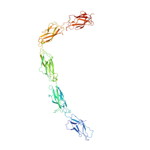
    Desmosomes are intercellular adhesive junctions that impart strength to vertebrate tissues. Their dense, ordered intercellular attachments are formed by desmogleins (Dsgs) and desmocollins (Dscs), but the nature of trans-cellular interactions between these specialized cadherins is unclear. Here, using solution biophysics and coated-bead aggregation experiments, we demonstrate family-wise heterophilic specificity: All Dsgs form adhesive dimers with all Dscs, with affinities characteristic of each Dsg:Dsc pair. Crystal structures of ectodomains from Dsg2 and Dsg3 and from Dsc1 and Dsc2 show binding through a strand-swap mechanism similar to that of homophilic classical cadherins. However, conserved charged amino acids inhibit Dsg:Dsg and Dsc:Dsc interactions by same-charge repulsion and promote heterophilic Dsg:Dsc interactions through opposite-charge attraction. These findings show that Dsg:Dsc heterodimers represent the fundamental adhesive unit of desmosomes and provide a structural framework for understanding desmosome assembly.


  • Organizational Affiliation

    Department of Systems Biology, Columbia University, New York, NY 10032; Howard Hughes Medical Institute, Columbia University, New York, NY 10032;


Macromolecules
Find similar proteins by:  (by identity cutoff)  |  3D Structure
Entity ID: 1
MoleculeChains Sequence LengthOrganismDetailsImage
Desmocollin-2,Desmoglein-2557Homo sapiensMutation(s): 0 
Gene Names: DSC2CDHF2DSC3DSG2CDHF5
UniProt & NIH Common Fund Data Resources
Find proteins for Q14126 (Homo sapiens)
Explore Q14126 
Go to UniProtKB:  Q14126
PHAROS:  Q14126
GTEx:  ENSG00000046604 
Find proteins for Q02487 (Homo sapiens)
Explore Q02487 
Go to UniProtKB:  Q02487
PHAROS:  Q02487
GTEx:  ENSG00000134755 
Entity Groups  
Sequence Clusters30% Identity50% Identity70% Identity90% Identity95% Identity100% Identity
UniProt GroupsQ14126Q02487
Sequence Annotations
Expand
  • Reference Sequence
Oligosaccharides

Help

Entity ID: 2
MoleculeChains Length2D Diagram Glycosylation3D Interactions
beta-D-galactopyranose-(1-4)-2-acetamido-2-deoxy-beta-D-glucopyranose-(1-3)-alpha-D-mannopyranose-(1-6)-[2-acetamido-2-deoxy-beta-D-glucopyranose-(1-2)-alpha-D-mannopyranose-(1-3)]beta-D-mannopyranose-(1-4)-2-acetamido-2-deoxy-beta-D-glucopyranose-(1-4)-2-acetamido-2-deoxy-beta-D-glucopyranose
B
8N-Glycosylation
Glycosylation Resources
GlyTouCan:  G88310KZ
GlyCosmos:  G88310KZ
GlyGen:  G88310KZ
Entity ID: 3
MoleculeChains Length2D Diagram Glycosylation3D Interactions
alpha-D-mannopyranose-(1-3)-[alpha-D-mannopyranose-(1-6)]beta-D-mannopyranose-(1-4)-2-acetamido-2-deoxy-beta-D-glucopyranose-(1-4)-[alpha-L-fucopyranose-(1-6)]2-acetamido-2-deoxy-beta-D-glucopyranose
C
6N-Glycosylation
Glycosylation Resources
GlyTouCan:  G82348BZ
GlyCosmos:  G82348BZ
GlyGen:  G82348BZ
Entity ID: 4
MoleculeChains Length2D Diagram Glycosylation3D Interactions
2-acetamido-2-deoxy-beta-D-glucopyranose-(1-4)-2-acetamido-2-deoxy-beta-D-glucopyranose
D
2N-Glycosylation
Glycosylation Resources
GlyTouCan:  G42666HT
GlyCosmos:  G42666HT
GlyGen:  G42666HT
Small Molecules
Ligands 4 Unique
IDChains Name / Formula / InChI Key2D Diagram3D Interactions
NAG
Query on NAG

Download Ideal Coordinates CCD File 
H [auth A],
I [auth A]
2-acetamido-2-deoxy-beta-D-glucopyranose
C8 H15 N O6
OVRNDRQMDRJTHS-FMDGEEDCSA-N
MAN
Query on MAN

Download Ideal Coordinates CCD File 
E [auth A],
F [auth A],
G [auth A]
alpha-D-mannopyranose
C6 H12 O6
WQZGKKKJIJFFOK-PQMKYFCFSA-N
CA
Query on CA

Download Ideal Coordinates CCD File 
J [auth A]
K [auth A]
L [auth A]
M [auth A]
N [auth A]
J [auth A],
K [auth A],
L [auth A],
M [auth A],
N [auth A],
O [auth A],
P [auth A],
Q [auth A],
R [auth A],
S [auth A],
T [auth A]
CALCIUM ION
Ca
BHPQYMZQTOCNFJ-UHFFFAOYSA-N
CL
Query on CL

Download Ideal Coordinates CCD File 
U [auth A],
V [auth A]
CHLORIDE ION
Cl
VEXZGXHMUGYJMC-UHFFFAOYSA-M
Experimental Data & Validation

Experimental Data

  • Method: X-RAY DIFFRACTION
  • Resolution: 3.29 Å
  • R-Value Free: 0.285 
  • R-Value Work: 0.247 
  • R-Value Observed: 0.249 
  • Space Group: P 2 21 21
Unit Cell:
Length ( Å )Angle ( ˚ )
a = 63.494α = 90
b = 87.489β = 90
c = 218.536γ = 90
Software Package:
Software NamePurpose
PHENIXrefinement
XDSdata reduction
XDSdata scaling
PHASERphasing

Structure Validation

View Full Validation Report


Ligand Structure Quality Assessment 


Entry History & Funding Information

Deposition Data


Funding OrganizationLocationGrant Number
National Institutes of Health/National Institute of General Medical Sciences (NIH/NIGMS)United StatesR01 GM062270

Revision History  (Full details and data files)

  • Version 1.0: 2016-06-22
    Type: Initial release
  • Version 1.1: 2016-06-29
    Changes: Database references
  • Version 1.2: 2016-07-13
    Changes: Database references
  • Version 1.3: 2017-09-20
    Changes: Author supporting evidence, Database references, Derived calculations
  • Version 1.4: 2019-12-25
    Changes: Author supporting evidence, Data collection
  • Version 2.0: 2020-07-29
    Type: Remediation
    Reason: Carbohydrate remediation
    Changes: Advisory, Atomic model, Data collection, Derived calculations, Structure summary
  • Version 2.1: 2023-09-27
    Changes: Data collection, Database references, Derived calculations, Refinement description, Structure summary