Replacing Arginine 33 for Alanine in the Hemophore HasA from Pseudomonas aeruginosa Causes Closure of the H32 Loop in the Apo-Protein.
Kumar, R., Qi, Y., Matsumura, H., Lovell, S., Yao, H., Battaile, K.P., Im, W., Moenne-Loccoz, P., Rivera, M.(2016) Biochemistry 55: 2622-2631
- PubMed: 27074415
- DOI: https://doi.org/10.1021/acs.biochem.6b00239
- Primary Citation of Related Structures:
5IQW, 5IQX - PubMed Abstract:
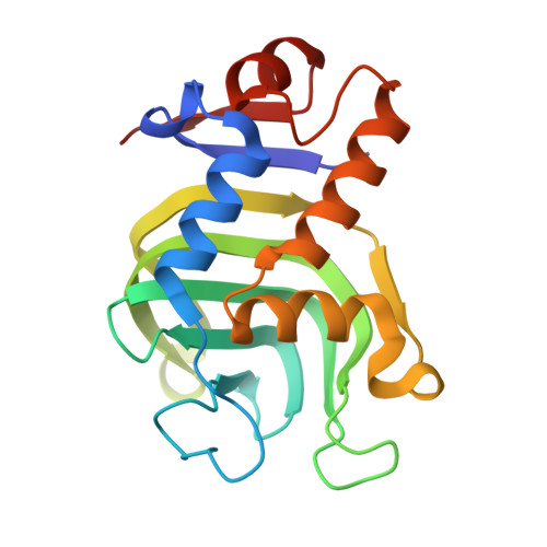
Previous characterization of hemophores from Serratia marcescens (HasAs), Pseudomonas aeruginosa (HasAp), and Yersinia pestis (HasAyp) showed that hemin binds between two loops, where it is axially coordinated by H32 and Y75. The Y75 loop is structurally conserved in all three hemophores and harbors conserved ligand Y75. The other loop contains H32 in HasAs and HasAp, but a noncoordinating Q32 in HasAyp. The H32 loop in apo-HasAs and apo-HasAp is in an open conformation, which places H32 about 30 Å from the hemin-binding site. Hence, hemin binding onto the Y75 loop of HasAs or HasAp triggers a large relocation of the H32 loop from an open- to a closed-loop conformation and enables coordination of the hemin-iron by H32. In comparison, the Q32 loop in apo-HasAyp is in the closed conformation, and hemin binding occurs with minimal reorganization and without coordinative interactions with the Q32 loop. Studies in crystallo and in solution have established that the open H32 loop in apo-HasAp and apo-HasAs is well structured and minimally affected by conformational dynamics. In this study we address the intriguing issue of the stability of the H32 loop in apo-HasAp and how hemin binding triggers its relocation. We address this question with a combination of NMR spectroscopy, X-ray crystallography, and molecular dynamics simulations and find that R33 is critical to the stability of the open H32 loop. Replacing R33 with A causes the H32 loop in R33A apo-HasAp to adopt a conformation similar to that of holo-HasAp. Finally, stopped-flow absorption and resonance Raman analyses of hemin binding to apo-R33A HasAp indicate that the closed H32 loop slows down the insertion of the heme inside the binding pocket, presumably as it obstructs access to the hydrophobic platform on the Y75 loop, but accelerates the completion of the heme iron coordination.
Organizational Affiliation:
Division of Environmental & Biomolecular Systems, Institute of Environmental Health, Oregon Health and Science University , 3181 SW Sam Jackson Park Road, Oregon 97239, United States.