5HS2

Crystal structure of IspD complexed with CTP and Mg2+ from Bacillus subtilis at 1.90 Angstroms resolution


Experimental Data Snapshot

  • Method: X-RAY DIFFRACTION
  • Resolution: 1.90 Å
  • R-Value Free: 0.252 
  • R-Value Work: 0.204 
  • R-Value Observed: 0.207 

wwPDB Validation   3D Report Full Report


Ligand Structure Quality Assessment 


This is version 1.1 of the entry. See complete history


Literature

A structural and functional study on the 2-C-methyl-d-erythritol-4-phosphate cytidyltransferase (IspD) from Bacillus subtilis.

Jin, Y.Liu, Z.Li, Y.Liu, W.Tao, Y.Wang, G.

(2016) Sci Rep 6: 36379-36379

  • DOI: https://doi.org/10.1038/srep36379
  • Primary Citation of Related Structures:  
    5DDT, 5DDV, 5HS2

  • PubMed Abstract: 

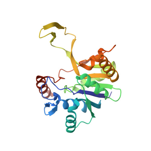
    2-C-Methyl-D-erythritol-4-phosphate cytidyltransferase (IspD) is an essential enzyme in the mevalonate-independent pathway of isoprenoid biosynthesis. This enzyme catalyzes 2-C-Methyl-d-erythritol 4-phosphate (MEP) and cytosine triphosphate (CTP) to 4-diphosphocytidyl-2-C-methyl-d-erythritol (CDPME) and inorganic pyrophosphate (PPi). Bacillus subtilis was a kind of excellent isoprene producer. However, the studies on the key enzymes of MEP pathway in B. subtilis were still absent. In this work, the crystal structures of IspD and IspD complexed with CTP from B.subtilis were determined. For the first time, the intact P-loop was observed in the apo structure of IspD enzyme. Structural comparisons revealed that the concerted movements of the P-loop and loops close to the active site were essential in the reaction catalyzed by IspD. Meanwhile, kinetic analysis showed that the CTP hydrolytic activity of IspD from B.subtilis was over two times higher than that from Escherichia coli. These results will be useful for future target-based screening of potential inhibitors and the metabolic engineering for isoprenoid biosynthesis.


  • Organizational Affiliation

    Key Laboratory of Environmental and Applied Microbiology, Chengdu Institute of Biology, Chinese Academy of Sciences, Chengdu, 610041, China.


Macromolecules
Find similar proteins by:  (by identity cutoff)  |  3D Structure
Entity ID: 1
MoleculeChains Sequence LengthOrganismDetailsImage
2-C-methyl-D-erythritol 4-phosphate cytidylyltransferase
A, B
232Bacillus subtilis subsp. subtilis str. 168Mutation(s): 0 
Gene Names: ispDyacMBSU00900
EC: 2.7.7.60
UniProt
Find proteins for Q06755 (Bacillus subtilis (strain 168))
Explore Q06755 
Go to UniProtKB:  Q06755
Entity Groups  
Sequence Clusters30% Identity50% Identity70% Identity90% Identity95% Identity100% Identity
UniProt GroupQ06755
Sequence Annotations
Expand
  • Reference Sequence
Small Molecules
Ligands 2 Unique
IDChains Name / Formula / InChI Key2D Diagram3D Interactions
CTP
Query on CTP

Download Ideal Coordinates CCD File 
C [auth A]CYTIDINE-5'-TRIPHOSPHATE
C9 H16 N3 O14 P3
PCDQPRRSZKQHHS-XVFCMESISA-N
MG
Query on MG

Download Ideal Coordinates CCD File 
D [auth A]MAGNESIUM ION
Mg
JLVVSXFLKOJNIY-UHFFFAOYSA-N
Experimental Data & Validation

Experimental Data

  • Method: X-RAY DIFFRACTION
  • Resolution: 1.90 Å
  • R-Value Free: 0.252 
  • R-Value Work: 0.204 
  • R-Value Observed: 0.207 
  • Space Group: P 1 21 1
Unit Cell:
Length ( Å )Angle ( ˚ )
a = 65.45α = 90
b = 49.43β = 101.41
c = 75.373γ = 90
Software Package:
Software NamePurpose
REFMACrefinement
HKL-2000data reduction
HKL-2000data scaling
PHASERphasing

Structure Validation

View Full Validation Report


Ligand Structure Quality Assessment 


Entry History 

Deposition Data

Revision History  (Full details and data files)

  • Version 1.0: 2016-11-23
    Type: Initial release
  • Version 1.1: 2023-11-08
    Changes: Data collection, Database references, Derived calculations, Refinement description