5EUH

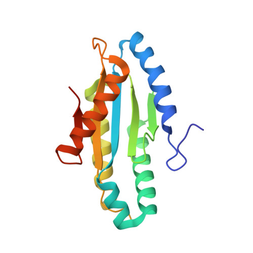
Crystal structure of the c-di-GMP-bound GGDEF domain of P. fluorescens GcbC

  • Classification: MEMBRANE PROTEIN
  • Organism(s): Pseudomonas fluorescens Pf0-1
  • Expression System: Escherichia coli
  • Mutation(s): No 

  • Deposited: 2015-11-18 Released: 2015-12-30 
  • Deposition Author(s): Giglio, K.M., Cooley, R.B., Sondermann, H.
  • Funding Organization(s): National Institutes of Health/National Institute Of Allergy and Infectious Diseases (NIH/NIAID), National Institutes of Health/National Institute of General Medical Sciences (NIH/NIGMS), National Science Foundation (NSF, United States)

Experimental Data Snapshot

  • Method: X-RAY DIFFRACTION
  • Resolution: 2.99 Å
  • R-Value Free: 0.243 
  • R-Value Work: 0.226 
  • R-Value Observed: 0.227 

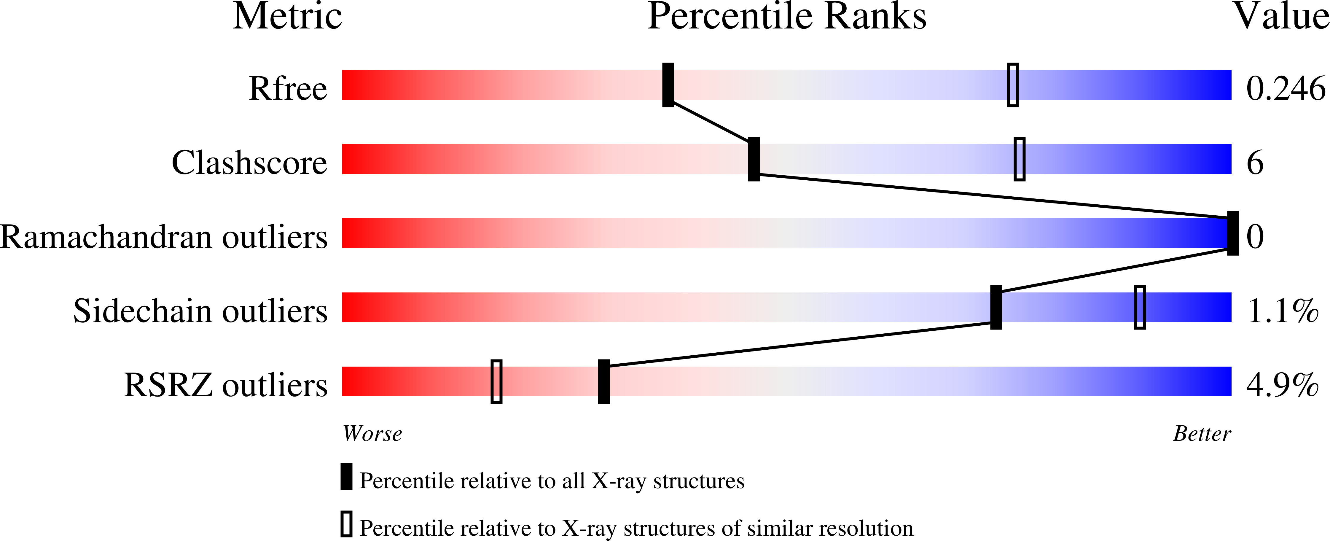
wwPDB Validation   3D Report Full Report


Ligand Structure Quality Assessment 


This is version 1.4 of the entry. See complete history


Literature

Contribution of Physical Interactions to Signaling Specificity between a Diguanylate Cyclase and Its Effector.

Dahlstrom, K.M.Giglio, K.M.Collins, A.J.Sondermann, H.O'Toole, G.A.

(2015) mBio 6: e01978-e01915

  • DOI: https://doi.org/10.1128/mBio.01978-15
  • Primary Citation of Related Structures:  
    5EUH

  • PubMed Abstract: 

    Cyclic diguanylate (c-di-GMP) is a bacterial second messenger that controls multiple cellular processes. c-di-GMP networks have up to dozens of diguanylate cyclases (DGCs) that synthesize c-di-GMP along with many c-di-GMP-responsive target proteins that can bind and respond to this signal. For such networks to have order, a mechanism(s) likely exists that allow DGCs to specifically signal their targets, and it has been suggested that physical interactions might provide such specificity. Our results show a DGC from Pseudomonas fluorescens physically interacting with its target protein at a conserved interface, and this interface can be predictive of DGC-target protein interactions. Furthermore, we demonstrate that physical interaction is necessary for the DGC to maximally signal its target. If such "local signaling" is a theme for even a fraction of the DGCs used by bacteria, it becomes possible to posit a model whereby physical interaction allows a DGC to directly signal its target protein, which in turn may help curtail undesired cross talk with other members of the network. An important question in microbiology is how bacteria make decisions using a signaling network made up of proteins that make, break, and bind the second messenger c-di-GMP, which is responsible for controlling many cellular behaviors. Previous work has shown that a given DGC enzyme will signal for specific cellular outputs, despite making the same diffusible molecule as its sibling DGCs in the unpartitioned space of the bacterial cell. Understanding how one DGC differentiates its output from the dozens of other such enzymes in the cell is synonymous with understanding a large component of the bacterial decision-making machinery. We present evidence for a helix on a DGC used to physically associate with its target protein, which is necessary to achieve maximal signaling.


  • Organizational Affiliation

    Department of Microbiology and Immunology, Geisel School of Medicine at Dartmouth, Hanover, New Hampshire.


Macromolecules
Find similar proteins by:  (by identity cutoff)  |  3D Structure
Entity ID: 1
MoleculeChains Sequence LengthOrganismDetailsImage
Putative GGDEF domain membrane protein
A, B, C, D
172Pseudomonas fluorescens Pf0-1Mutation(s): 0 
Gene Names: Pfl01_4666
UniProt
Find proteins for Q3K751 (Pseudomonas fluorescens (strain Pf0-1))
Explore Q3K751 
Go to UniProtKB:  Q3K751
Entity Groups  
Sequence Clusters30% Identity50% Identity70% Identity90% Identity95% Identity100% Identity
UniProt GroupQ3K751
Sequence Annotations
Expand
  • Reference Sequence
Small Molecules
Ligands 2 Unique
IDChains Name / Formula / InChI Key2D Diagram3D Interactions
C2E
Query on C2E

Download Ideal Coordinates CCD File 
E [auth A],
H [auth B],
I [auth B],
M [auth C]
9,9'-[(2R,3R,3aS,5S,7aR,9R,10R,10aS,12S,14aR)-3,5,10,12-tetrahydroxy-5,12-dioxidooctahydro-2H,7H-difuro[3,2-d:3',2'-j][1,3,7,9,2,8]tetraoxadiphosphacyclododecine-2,9-diyl]bis(2-amino-1,9-dihydro-6H-purin-6-one)
C20 H24 N10 O14 P2
PKFDLKSEZWEFGL-MHARETSRSA-N
SO4
Query on SO4

Download Ideal Coordinates CCD File 
F [auth A],
G [auth A],
J [auth B],
K [auth B],
L [auth B]
SULFATE ION
O4 S
QAOWNCQODCNURD-UHFFFAOYSA-L
Experimental Data & Validation

Experimental Data

  • Method: X-RAY DIFFRACTION
  • Resolution: 2.99 Å
  • R-Value Free: 0.243 
  • R-Value Work: 0.226 
  • R-Value Observed: 0.227 
  • Space Group: P 62
Unit Cell:
Length ( Å )Angle ( ˚ )
a = 141.913α = 90
b = 141.913β = 90
c = 106.169γ = 120
Software Package:
Software NamePurpose
PHENIXrefinement
HKL-2000data reduction
HKL-2000data scaling
PHENIXphasing

Structure Validation

View Full Validation Report


Ligand Structure Quality Assessment 


Entry History & Funding Information

Deposition Data


Funding OrganizationLocationGrant Number
National Institutes of Health/National Institute Of Allergy and Infectious Diseases (NIH/NIAID)United StatesR01-AI097307
National Institutes of Health/National Institute of General Medical Sciences (NIH/NIGMS)United StatesGM-103485
National Science Foundation (NSF, United States)United StatesDMR-1332208

Revision History  (Full details and data files)

  • Version 1.0: 2015-12-30
    Type: Initial release
  • Version 1.1: 2016-02-10
    Changes: Database references
  • Version 1.2: 2017-09-27
    Changes: Author supporting evidence, Derived calculations
  • Version 1.3: 2019-11-27
    Changes: Author supporting evidence
  • Version 1.4: 2023-09-27
    Changes: Data collection, Database references, Refinement description, Structure summary