5DG1

Sugar binding protein - human galectin-2


Experimental Data Snapshot

  • Method: X-RAY DIFFRACTION
  • Resolution: 3.20 Å
  • R-Value Free: 0.321 
  • R-Value Work: 0.297 
  • R-Value Observed: 0.300 

wwPDB Validation   3D Report Full Report


This is version 2.1 of the entry. See complete history


Literature

Human galectin-2 interacts with carbohydrates and peptides non-classically: new insight from X-ray crystallography and hemagglutination.

Si, Y.Feng, S.Gao, J.Wang, Y.Zhang, Z.Meng, Y.Zhou, Y.Tai, G.Su, J.

(2016) Acta Biochim Biophys Sin (Shanghai) 

  • DOI: https://doi.org/10.1093/abbs/gmw089
  • Primary Citation of Related Structures:  
    5DG1, 5DG2, 5EWS

  • PubMed Abstract: 

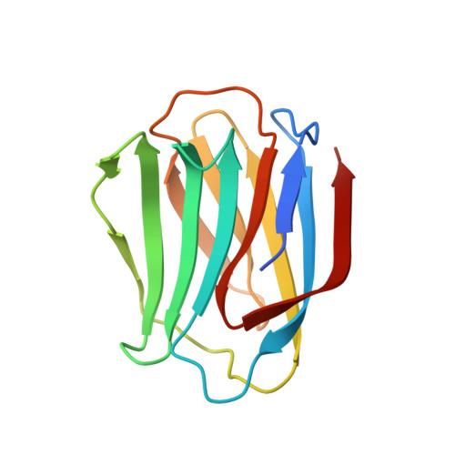
    Galectin-2 (Gal-2) plays a role in cancer, myocardial infarction, immune response, and gastrointestinal tract diseases. The only reported crystal structure of Gal-2 shows that it is a dimer in which the monomer subunits have almost identical structures, each binding with one molecule of lactose. In this study, we crystallized Gal-2 under new conditions that produced three crystal structures. In each Gal-2 dimer structure, lactose was shown to be bound to only one of the carbohydrate recognition domain subunits. In solution studies, the thermal shift assay demonstrated that inequivalent monomer subunits in the Gal-2 dimer become equivalent upon ligand binding. In addition, galectin-mediated erythrocyte agglutination assays using lactose and larger complex polysaccharides as inhibitors showed the structural differences between Gal-1 and Gal-2. Overall, our results reveal some novel aspects to the structural differentiation in Gal-2 and expand the potential for different types of molecular interactions that may be specific to this lectin.


  • Organizational Affiliation

    Jilin Province Key Laboratory on Chemistry and Biology of Natural Drugs in Changbai Mountain, School of Life Sciences, Northeast Normal University, Changchun 130024, China.


Macromolecules
Find similar proteins by:  (by identity cutoff)  |  3D Structure
Entity ID: 1
MoleculeChains Sequence LengthOrganismDetailsImage
Galectin-2135Homo sapiensMutation(s): 0 
Gene Names: LGALS2
UniProt & NIH Common Fund Data Resources
Find proteins for P05162 (Homo sapiens)
Explore P05162 
Go to UniProtKB:  P05162
PHAROS:  P05162
GTEx:  ENSG00000100079 
Entity Groups  
Sequence Clusters30% Identity50% Identity70% Identity90% Identity95% Identity100% Identity
UniProt GroupP05162
Sequence Annotations
Expand
  • Reference Sequence
Oligosaccharides

Help

Entity ID: 2
MoleculeChains Length2D Diagram Glycosylation3D Interactions
beta-D-galactopyranose-(1-4)-beta-D-glucopyranoseG [auth E],
H [auth F],
I [auth G]
2N/A
Glycosylation Resources
GlyTouCan:  G84224TW
GlyCosmos:  G84224TW
GlyGen:  G84224TW
Biologically Interesting Molecules (External Reference) 1 Unique
Experimental Data & Validation

Experimental Data

  • Method: X-RAY DIFFRACTION
  • Resolution: 3.20 Å
  • R-Value Free: 0.321 
  • R-Value Work: 0.297 
  • R-Value Observed: 0.300 
  • Space Group: P 2 21 21
Unit Cell:
Length ( Å )Angle ( ˚ )
a = 38.5α = 90
b = 106.78β = 90
c = 182.11γ = 90
Software Package:
Software NamePurpose
PHENIXrefinement
PDB_EXTRACTdata extraction

Structure Validation

View Full Validation Report


Entry History 

Deposition Data

Revision History  (Full details and data files)

  • Version 1.0: 2016-09-14
    Type: Initial release
  • Version 1.1: 2017-09-27
    Changes: Data collection, Derived calculations
  • Version 2.0: 2020-07-29
    Type: Remediation
    Reason: Carbohydrate remediation
    Changes: Advisory, Atomic model, Data collection, Derived calculations, Structure summary
  • Version 2.1: 2023-11-08
    Changes: Data collection, Database references, Derived calculations, Refinement description, Structure summary