5CNW

Crystal structure of a novel disulfide oxidoreductase from Deinococcus radiodurans


Experimental Data Snapshot

  • Method: X-RAY DIFFRACTION
  • Resolution: 1.65 Å
  • R-Value Free: 0.198 
  • R-Value Work: 0.170 
  • R-Value Observed: 0.172 

wwPDB Validation   3D Report Full Report


This is version 1.3 of the entry. See complete history


Literature

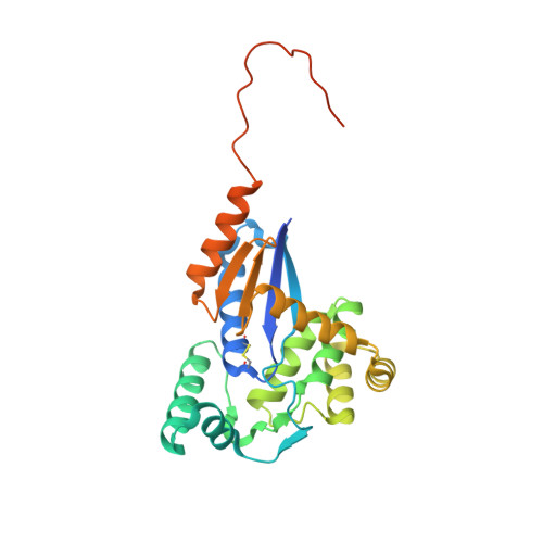
drFrnE Represents a Hitherto Unknown Class of Eubacterial Cytoplasmic Disulfide Oxido-Reductases.

Bihani, S.C.Panicker, L.Rajpurohit, Y.S.Misra, H.S.Kumar, V.

(2018) Antioxid Redox Signal 28: 296-310

  • DOI: https://doi.org/10.1089/ars.2016.6960
  • Primary Citation of Related Structures:  
    5CNW, 5CO3, 5COH, 5E59

  • PubMed Abstract: 

    Living cells employ thioredoxin and glutaredoxin disulfide oxido-reductases to protect thiol groups in intracellular proteins. FrnE protein of Deinococcus radiodurans (drFrnE) is a disulfide oxido-reductase that is induced in response to Cd 2+ exposure and is involved in cadmium and radiation tolerance. The aim of this study is to probe structure, function, and cellular localization of FrnE class of proteins.


  • Organizational Affiliation

    1 Protein Crystallography Section, Radiation Biology & Health Sciences Division, Bhabha Atomic Research Centre , Mumbai, India .


Macromolecules
Find similar proteins by:  (by identity cutoff)  |  3D Structure
Entity ID: 1
MoleculeChains Sequence LengthOrganismDetailsImage
FrnE protein260Deinococcus radiodurans R1 = ATCC 13939 = DSM 20539Mutation(s): 0 
Gene Names: DR_0659
UniProt
Find proteins for Q9RWK7 (Deinococcus radiodurans (strain ATCC 13939 / DSM 20539 / JCM 16871 / CCUG 27074 / LMG 4051 / NBRC 15346 / NCIMB 9279 / VKM B-1422 / R1))
Explore Q9RWK7 
Go to UniProtKB:  Q9RWK7
Entity Groups  
Sequence Clusters30% Identity50% Identity70% Identity90% Identity95% Identity100% Identity
UniProt GroupQ9RWK7
Sequence Annotations
Expand
  • Reference Sequence
Small Molecules
Ligands 1 Unique
IDChains Name / Formula / InChI Key2D Diagram3D Interactions
GOL
Query on GOL

Download Ideal Coordinates CCD File 
B [auth A]GLYCEROL
C3 H8 O3
PEDCQBHIVMGVHV-UHFFFAOYSA-N
Experimental Data & Validation

Experimental Data

  • Method: X-RAY DIFFRACTION
  • Resolution: 1.65 Å
  • R-Value Free: 0.198 
  • R-Value Work: 0.170 
  • R-Value Observed: 0.172 
  • Space Group: P 21 2 21
Unit Cell:
Length ( Å )Angle ( ˚ )
a = 47.805α = 90
b = 63.032β = 90
c = 87.298γ = 90
Software Package:
Software NamePurpose
PHENIXrefinement
PDB_EXTRACTdata extraction
XDSdata processing
Aimlessdata reduction
PHASERphasing
XDSdata scaling

Structure Validation

View Full Validation Report


Entry History 

Deposition Data

Revision History  (Full details and data files)

  • Version 1.0: 2016-07-20
    Type: Initial release
  • Version 1.1: 2018-07-04
    Changes: Data collection, Database references, Derived calculations
  • Version 1.2: 2019-04-10
    Changes: Data collection, Database references
  • Version 1.3: 2023-11-08
    Changes: Data collection, Database references, Refinement description