5B0Q

beta-1,2-Mannobiose phosphorylase from Listeria innocua - mannose complex


Experimental Data Snapshot

  • Method: X-RAY DIFFRACTION
  • Resolution: 2.30 Å
  • R-Value Free: 0.217 
  • R-Value Work: 0.172 
  • R-Value Observed: 0.174 

Starting Model: experimental
View more details

wwPDB Validation   3D Report Full Report


Ligand Structure Quality Assessment 


This is version 1.4 of the entry. See complete history


Literature

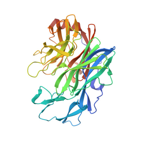
Characterization and crystal structure determination of beta-1,2-mannobiose phosphorylase from Listeria innocua

Tsuda, T.Nihira, T.Chiku, K.Suzuki, E.Arakawa, T.Nishimoto, M.Kitaoka, M.Nakai, H.Fushinobu, S.

(2015) FEBS Lett 589: 3816-3821

  • DOI: https://doi.org/10.1016/j.febslet.2015.11.034
  • Primary Citation of Related Structures:  
    5B0P, 5B0Q, 5B0R, 5B0S

  • PubMed Abstract: 

    Glycoside hydrolase family 130 consists of phosphorylases and hydrolases for β-mannosides. Here, we characterized β-1,2-mannobiose phosphorylase from Listeria innocua (Lin0857) and determined its crystal structures complexed with β-1,2-linked mannooligosaccharides. β-1,2-Mannotriose was bound in a U-shape, interacting with a phosphate analog at both ends. Lin0857 has a unique dimer structure connected by a loop, and a significant open-close loop displacement was observed for substrate entry. A long loop, which is exclusively present in Lin0857, covers the active site to limit the pocket size. A structural basis for substrate recognition and phosphorolysis was provided.


  • Organizational Affiliation

    Department of Biotechnology, The University of Tokyo, 1-1-1 Yayoi, Bunkyo-ku, Tokyo 113-8657, Japan.


Macromolecules
Find similar proteins by:  (by identity cutoff)  |  3D Structure
Entity ID: 1
MoleculeChains Sequence LengthOrganismDetailsImage
Lin0857 protein
A, B
363Listeria innocua Clip11262Mutation(s): 0 
Gene Names: lin0857
UniProt
Find proteins for Q92DF6 (Listeria innocua serovar 6a (strain ATCC BAA-680 / CLIP 11262))
Explore Q92DF6 
Go to UniProtKB:  Q92DF6
Entity Groups  
Sequence Clusters30% Identity50% Identity70% Identity90% Identity95% Identity100% Identity
UniProt GroupQ92DF6
Sequence Annotations
Expand
  • Reference Sequence
Experimental Data & Validation

Experimental Data

  • Method: X-RAY DIFFRACTION
  • Resolution: 2.30 Å
  • R-Value Free: 0.217 
  • R-Value Work: 0.172 
  • R-Value Observed: 0.174 
  • Space Group: P 31 2 1
Unit Cell:
Length ( Å )Angle ( ˚ )
a = 146.212α = 90
b = 146.212β = 90
c = 105.841γ = 120
Software Package:
Software NamePurpose
REFMACrefinement
HKL-2000data reduction
HKL-2000data scaling
MOLREPphasing

Structure Validation

View Full Validation Report


Ligand Structure Quality Assessment 


Entry History & Funding Information

Deposition Data


Funding OrganizationLocationGrant Number
The Ministry of Agriculture, Forestry and Fisheries, JapanJapan25010A

Revision History  (Full details and data files)

  • Version 1.0: 2015-12-02
    Type: Initial release
  • Version 1.1: 2015-12-23
    Changes: Database references
  • Version 1.2: 2020-02-26
    Changes: Data collection, Database references, Derived calculations
  • Version 1.3: 2020-07-29
    Type: Remediation
    Reason: Carbohydrate remediation
    Changes: Data collection, Derived calculations, Structure summary
  • Version 1.4: 2023-11-08
    Changes: Data collection, Database references, Refinement description, Structure summary