5AZH

Crystal structure of LGG-2 fused with an EEEWEEL peptide


Experimental Data Snapshot

  • Method: X-RAY DIFFRACTION
  • Resolution: 2.30 Å
  • R-Value Free: 0.288 
  • R-Value Work: 0.236 
  • R-Value Observed: 0.238 

wwPDB Validation   3D Report Full Report


This is version 1.1 of the entry. See complete history


Literature

Structural Basis of the Differential Function of the Two C. elegans Atg8 Homologs, LGG-1 and LGG-2, in Autophagy.

Wu, F.Watanabe, Y.Guo, X.Y.Qi, X.Wang, P.Zhao, H.Y.Wang, Z.Fujioka, Y.Zhang, H.Ren, J.Q.Fang, T.C.Shen, Y.X.Feng, W.Hu, J.J.Noda, N.N.Zhang, H.

(2015) Mol Cell 60: 914-929

  • DOI: https://doi.org/10.1016/j.molcel.2015.11.019
  • Primary Citation of Related Structures:  
    5AZF, 5AZG, 5AZH, 5E6N, 5E6O

  • PubMed Abstract: 

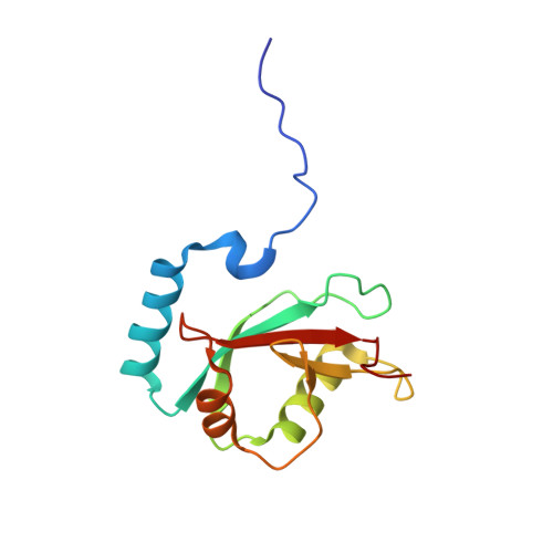
    Multicellular organisms have multiple homologs of the yeast ATG8 gene, but the differential roles of these homologs in autophagy during development remain largely unknown. Here we investigated structure/function relationships in the two C. elegans Atg8 homologs, LGG-1 and LGG-2. lgg-1 is essential for degradation of protein aggregates, while lgg-2 has cargo-specific and developmental-stage-specific roles in aggregate degradation. Crystallography revealed that the N-terminal tails of LGG-1 and LGG-2 adopt the closed and open form, respectively. LGG-1 and LGG-2 interact differentially with autophagy substrates and Atg proteins, many of which carry a LIR motif. LGG-1 and LGG-2 have structurally distinct substrate binding pockets that prefer different residues in the interacting LIR motif, thus influencing binding specificity. Lipidated LGG-1 and LGG-2 possess distinct membrane tethering and fusion activities, which may result from the N-terminal differences. Our study reveals the differential function of two ATG8 homologs in autophagy during C. elegans development.


  • Organizational Affiliation

    National Laboratory of Biomacromolecules, Institute of Biophysics, Chinese Academy of Sciences, Beijing 100101, P.R. China.


Macromolecules
Find similar proteins by:  (by identity cutoff)  |  3D Structure
Entity ID: 1
MoleculeChains Sequence LengthOrganismDetailsImage
EEEWEEL peptide,Protein lgg-2128synthetic constructCaenorhabditis elegans
This entity is chimeric
Mutation(s): 0 
UniProt
Find proteins for Q23536 (Caenorhabditis elegans)
Explore Q23536 
Go to UniProtKB:  Q23536
Entity Groups  
Sequence Clusters30% Identity50% Identity70% Identity90% Identity95% Identity100% Identity
UniProt GroupQ23536
Sequence Annotations
Expand
  • Reference Sequence
Small Molecules
Ligands 1 Unique
IDChains Name / Formula / InChI Key2D Diagram3D Interactions
MG
Query on MG

Download Ideal Coordinates CCD File 
B [auth A]MAGNESIUM ION
Mg
JLVVSXFLKOJNIY-UHFFFAOYSA-N
Experimental Data & Validation

Experimental Data

  • Method: X-RAY DIFFRACTION
  • Resolution: 2.30 Å
  • R-Value Free: 0.288 
  • R-Value Work: 0.236 
  • R-Value Observed: 0.238 
  • Space Group: P 21 21 21
Unit Cell:
Length ( Å )Angle ( ˚ )
a = 35.532α = 90
b = 55.193β = 90
c = 72.592γ = 90
Software Package:
Software NamePurpose
REFMACrefinement
HKL-2000data reduction
HKL-2000data scaling
Cootmodel building

Structure Validation

View Full Validation Report


Entry History & Funding Information

Deposition Data


Funding OrganizationLocationGrant Number
Ministry of Education, Culture, Sports, Science and Technology of JapanJapan25111004
Ministry of Education, Culture, Sports, Science and Technology of JapanJapan26870828
Japan Science and Technology AgencyJapanCREST

Revision History  (Full details and data files)

  • Version 1.0: 2015-12-30
    Type: Initial release
  • Version 1.1: 2020-01-01
    Changes: Data collection, Database references, Derived calculations