The N-Terminal Pre-A Region of Mycobacterium Tuberculosis 2/2Hbn Promotes No-Dioxygenase Activity.
Pesce, A., Bustamante, J.P., Bidon-Chanal, A., Boechi, L., Estrin, D.A., Luque, F.J., Sebilo, A., Guertin, M., Bolognesi, M., Ascenzi, P., Nardini, M.(2016) FEBS J 283: 305
- PubMed: 26499089
- DOI: https://doi.org/10.1111/febs.13571
- Primary Citation of Related Structures:
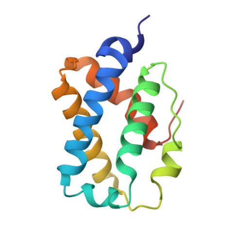
5AB8 - PubMed Abstract:
A unique defense mechanisms by which Mycobacterium tuberculosis protects itself from nitrosative stress is based on the O2 -dependent NO-dioxygenase (NOD) activity of truncated hemoglobin 2/2HbN (Mt2/2HbN). The NOD activity largely depends on the efficiency of ligand migration to the heme cavity through a two-tunnel (long and short) system; recently, it was also correlated with the presence at the Mt2/2HbN N-terminus of a short pre-A region, not conserved in most 2/2HbNs, whose deletion results in a drastic reduction of NO scavenging. In the present study, we report the crystal structure of Mt2/2HbN-ΔpreA, lacking the pre-A region, at a resolution of 1.53 Å. We show that removal of the pre-A region results in long range effects on the protein C-terminus, promoting the assembly of a stable dimer, both in the crystals and in solution. In the Mt2/2HbN-ΔpreA dimer, access of heme ligands to the short tunnel is hindered. Molecular dynamics simulations show that the long tunnel branch is the only accessible pathway for O2 -ligand migration to/from the heme, and that the gating residue Phe(62)E15 partly restricts the diameter of the tunnel. Accordingly, kinetic measurements indicate that the kon value for peroxynitrite isomerization by Mt2/2HbN-ΔpreA-Fe(III) is four-fold lower relative to the full-length protein, and that NO scavenging by Mt2/2HbN-ΔpreA-Fe(II)-O2 is reduced by 35-fold. Therefore, we speculate that Mt2/2HbN evolved to host the pre-A region as a mechanism for preventing dimerization, thus reinforcing the survival of the microorganism against the reactive nitrosative stress in macrophages. Coordinates and structure factors have been deposited in the Protein Data Bank under accession number 5AB8.
Organizational Affiliation:
Department of Physics, University of Genova, Italy.