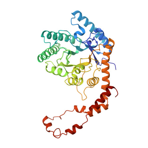
Structures of D-xylose isomerase from Arthrobacter strain B3728 containing the inhibitors xylitol and D-sorbitol at 2.5 A and 2.3 A resolution, respectively.
Henrick, K., Collyer, C.A., Blow, D.M.(1989) J Mol Biol 208: 129-157
- PubMed: 2769749
- DOI: https://doi.org/10.1016/0022-2836(89)90092-2
- Primary Citation of Related Structures:
4XIA, 5XIA - PubMed Abstract:
The structures of D-xylose isomerase from Arthrobacter strain B3728 containing the polyol inhibitors xylitol and D-sorbitol have been solved at 2.5 A and 2.3 A, respectively. The structures have been refined using restrained least-squares refinement methods. The final crystallographic R-factors for the D-sorbitol (xylitol) bound molecules, for 43,615 (32,989) reflections are 15.6 (14.7). The molecule is a tetramer and the asymmetric unit of the crystal contains a dimer, the final model of which, incorporates a total of 6086 unique protein, inhibitor and magnesium atoms together with 535 bound solvent molecules. Each subunit of the enzyme contains two domains: the main domain is a parallel-stranded alpha-beta barrel, which has been reported in 14 other enzymes. The C-terminal domain is a loop structure consisting of five helical segments and is involved in intermolecular contacts between subunits that make up the tetramer. The structures have been analysed with respect to molecular symmetry, intersubunit contacts, inhibitor binding and active site geometry. The refined model shows the two independent subunits to be similar apart from local deviations due to solvent contacts in the solvent-exposed helices. The enzyme is dependent on a divalent cation for catalytic activity. Two metal ions are required per monomer, and the high-affinity magnesium(II) site has been identified from the structural results presented here. The metal ion is complexed, at the high-affinity site, by four carboxylate side-chains of the conserved residues, Glu180, Glu216, Asp244 and Asp292. The inhibitor polyols are bound in the active site in an extended open chain conformation and complete an octahedral co-ordination shell for the magnesium cation via their oxygen atoms O-2 and O-4. The active site lies in a deep pocket near the C-terminal ends of the beta-strands of the barrel domain and includes residues from a second subunit. The tetrameric molecule can be considered to be a dimer of "active" dimers, the active sites being composed of residues from both subunits. The analysis has revealed the presence of several internal salt-bridges stabilizing the tertiary and quaternary structure. One of these, between Asp23 and Arg139, appears to play a key role in stabilizing the active dimer and is conserved in the known sequences of this enzyme.(ABSTRACT TRUNCATED AT 400 WORDS)
Organizational Affiliation:
Blackett Laboratory, Imperial College, London, England.