


Crystal structure of Leucine-, Isoleucine-, Valine-, Threonine-, and Alanine-binding protein from Brucella ovis
Abendroth, J., Fairman, J.W., Lorimer, D.D., Edwards, T.E.To be published.
Experimental Data Snapshot
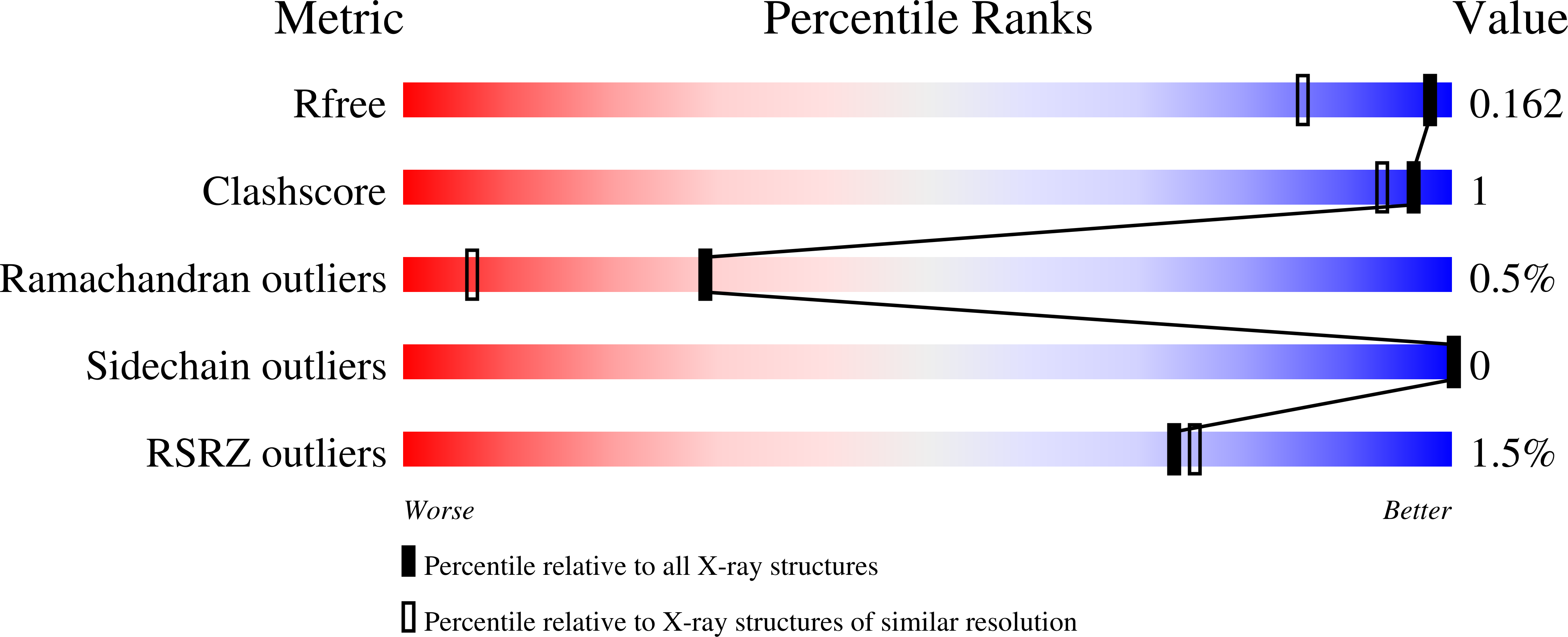
wwPDB Validation 3D Report Full Report
Entity ID: 1 | |||||
|---|---|---|---|---|---|
| Molecule | Chains | Sequence Length | Organism | Details | Image |
| Putative branched chain amino acid ABC transporter, periplasmic amino acid-binding protein | 399 | Brucella ovis ATCC 25840 | Mutation(s): 1 Gene Names: BOV_A0021 | 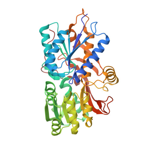 | |
UniProt | |||||
Find proteins for A0A0J9X260 (Brucella ovis (strain ATCC 25840 / 63/290 / NCTC 10512)) Explore A0A0J9X260 Go to UniProtKB: A0A0J9X260 | |||||
Entity Groups | |||||
| Sequence Clusters | 30% Identity50% Identity70% Identity90% Identity95% Identity100% Identity | ||||
| UniProt Group | A0A0J9X260 | ||||
Sequence AnnotationsExpand | |||||
| |||||
| Ligands 2 Unique | |||||
|---|---|---|---|---|---|
| ID | Chains | Name / Formula / InChI Key | 2D Diagram | 3D Interactions | |
| ACT Query on ACT | B [auth A], C [auth A] | ACETATE ION C2 H3 O2 QTBSBXVTEAMEQO-UHFFFAOYSA-M |  | ||
| NA Query on NA | D [auth A], E [auth A] | SODIUM ION Na FKNQFGJONOIPTF-UHFFFAOYSA-N |  | ||
| Length ( Å ) | Angle ( ˚ ) |
|---|---|
| a = 62.12 | α = 90 |
| b = 46.39 | β = 101.62 |
| c = 62.4 | γ = 90 |
| Software Name | Purpose |
|---|---|
| PHENIX | refinement |
| XDS | data reduction |
| XSCALE | data scaling |
| PHASER | phasing |
| ARP | model building |
| WARP | model building |
| Coot | model building |
| PDB_EXTRACT | data extraction |
RCSB PDB (citation) is hosted by
RCSB PDB is a member of the

