4WBQ

Crystal structure of the exonuclease domain of QIP (QDE-2 interacting protein) solved by native-SAD phasing.


Experimental Data Snapshot

  • Method: X-RAY DIFFRACTION
  • Resolution: 2.69 Å
  • R-Value Free: 0.227 
  • R-Value Work: 0.172 
  • R-Value Observed: 0.175 

wwPDB Validation   3D Report Full Report


This is version 1.2 of the entry. See complete history


Literature

Fast native-SAD phasing for routine macromolecular structure determination.

Weinert, T.Olieric, V.Waltersperger, S.Panepucci, E.Chen, L.Zhang, H.Zhou, D.Rose, J.Ebihara, A.Kuramitsu, S.Li, D.Howe, N.Schnapp, G.Pautsch, A.Bargsten, K.Prota, A.E.Surana, P.Kottur, J.Nair, D.T.Basilico, F.Cecatiello, V.Pasqualato, S.Boland, A.Weichenrieder, O.Wang, B.C.Steinmetz, M.O.Caffrey, M.Wang, M.

(2015) Nat Methods 12: 131-133

  • DOI: https://doi.org/10.1038/nmeth.3211
  • Primary Citation of Related Structures:  
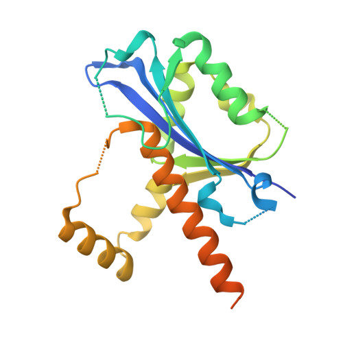
    4PGO, 4PII, 4R8T, 4R8U, 4TN8, 4TNO, 4WAB, 4WAU, 4WBQ, 4WBX

  • PubMed Abstract: 

    We describe a data collection method that uses a single crystal to solve X-ray structures by native SAD (single-wavelength anomalous diffraction). We solved the structures of 11 real-life examples, including a human membrane protein, a protein-DNA complex and a 266-kDa multiprotein-ligand complex, using this method. The data collection strategy is suitable for routine structure determination and can be implemented at most macromolecular crystallography synchrotron beamlines.


  • Organizational Affiliation

    Swiss Light Source at Paul Scherrer Institut, Villigen, Switzerland.


Macromolecules
Find similar proteins by:  (by identity cutoff)  |  3D Structure
Entity ID: 1
MoleculeChains Sequence LengthOrganismDetailsImage
QDE-2-interacting protein
A, B
240Neurospora crassaMutation(s): 0 
UniProt
Find proteins for A5JQJ3 (Neurospora crassa)
Explore A5JQJ3 
Go to UniProtKB:  A5JQJ3
Entity Groups  
Sequence Clusters30% Identity50% Identity70% Identity90% Identity95% Identity100% Identity
UniProt GroupA5JQJ3
Sequence Annotations
Expand
  • Reference Sequence
Experimental Data & Validation

Experimental Data

  • Method: X-RAY DIFFRACTION
  • Resolution: 2.69 Å
  • R-Value Free: 0.227 
  • R-Value Work: 0.172 
  • R-Value Observed: 0.175 
  • Space Group: P 32 2 1
Unit Cell:
Length ( Å )Angle ( ˚ )
a = 77.04α = 90
b = 77.04β = 90
c = 112.31γ = 120
Software Package:
Software NamePurpose
PHENIXrefinement
XDSdata reduction
XSCALEdata scaling
SHELXDEphasing

Structure Validation

View Full Validation Report


Entry History 

Deposition Data

Revision History  (Full details and data files)

  • Version 1.0: 2014-12-10
    Type: Initial release
  • Version 1.1: 2014-12-31
    Changes: Database references
  • Version 1.2: 2015-02-11
    Changes: Database references