4UD1

Structure of the N Terminal domain of the MERS CoV nucleocapsid


Experimental Data Snapshot

  • Method: X-RAY DIFFRACTION
  • Resolution: 2.48 Å
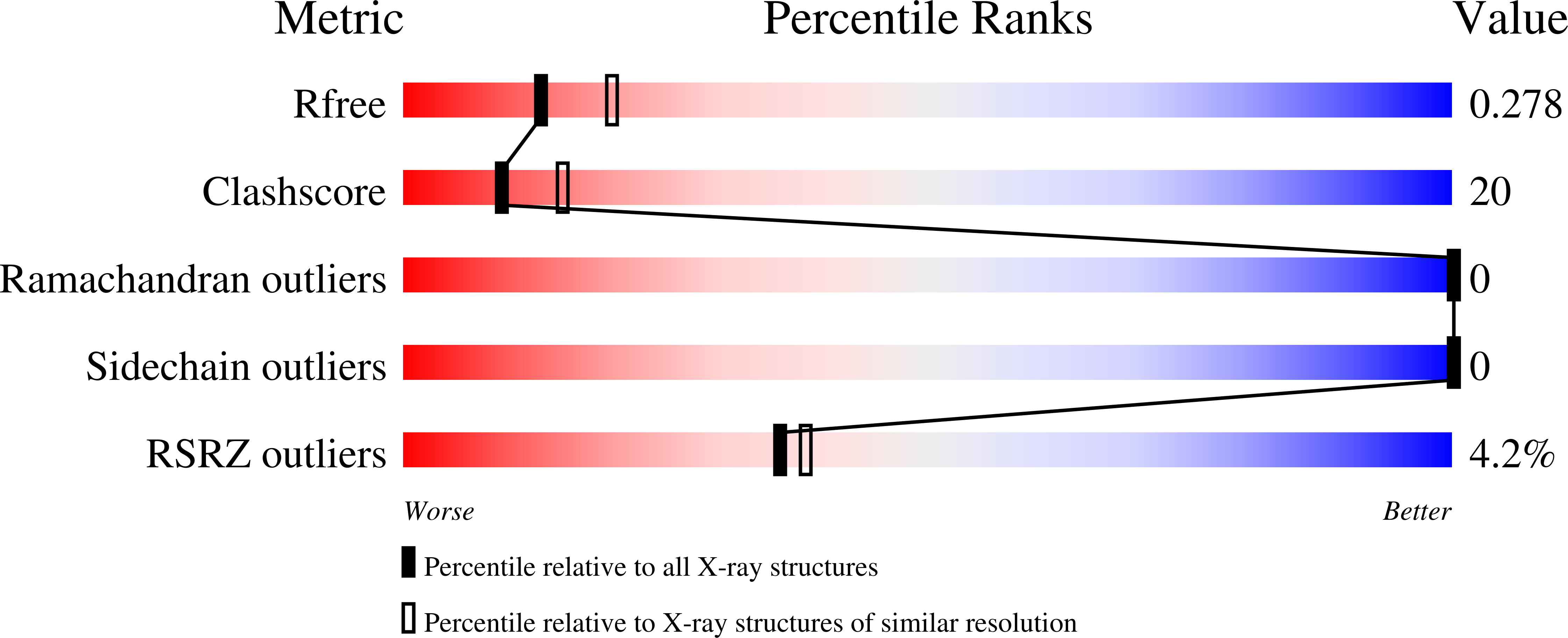
  • R-Value Free: 0.274 
  • R-Value Work: 0.228 
  • R-Value Observed: 0.231 

wwPDB Validation   3D Report Full Report


This is version 1.4 of the entry. See complete history


Literature

Structural Characterization of the N-Terminal Part of the Mers-Cov Nucleocapsid by X-Ray Diffraction and Small-Angle X-Ray Scattering

Papageorgiou, N.Lichiere, J.Baklouti, A.Ferron, F.Canard, B.Coutard, B.

(2016) Acta Crystallogr D Biol Crystallogr 72: 192

  • DOI: https://doi.org/10.1107/S2059798315024328
  • Primary Citation of Related Structures:  
    4UD1

  • PubMed Abstract: 

    The N protein of coronaviruses is a multifunctional protein that is organized into several domains. The N-terminal part is composed of an intrinsically disordered region (IDR) followed by a structured domain called the N-terminal domain (NTD). In this study, the structure determination of the N-terminal region of the MERS-CoV N protein via X-ray diffraction measurements is reported at a resolution of 2.4 Å. Since the first 30 amino acids were not resolved by X-ray diffraction, the structural study was completed by a SAXS experiment to propose a structural model including the IDR. This model presents the N-terminal region of the MERS-CoV as a monomer that displays structural features in common with other coronavirus NTDs.


  • Organizational Affiliation

    CNRS, AFMB UMR 7257, 13288 Marseille, France.


Macromolecules
Find similar proteins by:  (by identity cutoff)  |  3D Structure
Entity ID: 1
MoleculeChains Sequence LengthOrganismDetailsImage
N PROTEIN
A, B, C, D, E
164Middle East respiratory syndrome-related coronavirusMutation(s): 0 
UniProt
Find proteins for T2B9R0 (Middle East respiratory syndrome-related coronavirus)
Explore T2B9R0 
Go to UniProtKB:  T2B9R0
Entity Groups  
Sequence Clusters30% Identity50% Identity70% Identity90% Identity95% Identity100% Identity
UniProt GroupT2B9R0
Sequence Annotations
Expand
  • Reference Sequence
Small Molecules
Ligands 3 Unique
IDChains Name / Formula / InChI Key2D Diagram3D Interactions
GOL
Query on GOL

Download Ideal Coordinates CCD File 
I [auth B],
L [auth C],
M [auth D]
GLYCEROL
C3 H8 O3
PEDCQBHIVMGVHV-UHFFFAOYSA-N
IMD
Query on IMD

Download Ideal Coordinates CCD File 
H [auth B],
K [auth C]
IMIDAZOLE
C3 H5 N2
RAXXELZNTBOGNW-UHFFFAOYSA-O
NH4
Query on NH4

Download Ideal Coordinates CCD File 
F [auth A],
G [auth B],
J [auth C],
N [auth E]
AMMONIUM ION
H4 N
QGZKDVFQNNGYKY-UHFFFAOYSA-O
Experimental Data & Validation

Experimental Data

  • Method: X-RAY DIFFRACTION
  • Resolution: 2.48 Å
  • R-Value Free: 0.274 
  • R-Value Work: 0.228 
  • R-Value Observed: 0.231 
  • Space Group: C 1 2 1
Unit Cell:
Length ( Å )Angle ( ˚ )
a = 208.648α = 90
b = 67.021β = 89.98
c = 60.94γ = 90
Software Package:
Software NamePurpose
PHENIXrefinement
XDSdata reduction
PHENIXdata scaling
PHASERphasing

Structure Validation

View Full Validation Report


Entry History 

Deposition Data

Revision History  (Full details and data files)

  • Version 1.0: 2015-12-02
    Type: Initial release
  • Version 1.1: 2016-02-03
    Changes: Data collection
  • Version 1.2: 2016-02-10
    Changes: Database references
  • Version 1.3: 2016-03-02
    Changes: Database references
  • Version 1.4: 2023-12-20
    Changes: Data collection, Database references, Derived calculations, Other, Refinement description