4QQU

Crystal structure of the cobalamin-independent methionine synthase enzyme in a closed conformation


Experimental Data Snapshot

  • Method: X-RAY DIFFRACTION
  • Resolution: 2.98 Å
  • R-Value Free: 0.276 
  • R-Value Work: 0.175 
  • R-Value Observed: 0.180 

wwPDB Validation   3D Report Full Report


Ligand Structure Quality Assessment 


This is version 1.4 of the entry. See complete history


Literature

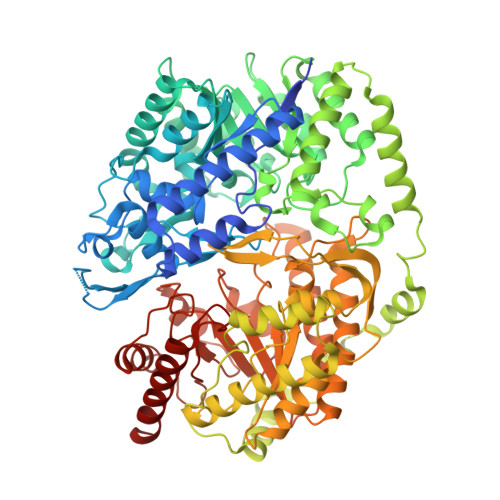
The cobalamin-independent methionine synthase enzyme captured in a substrate-induced closed conformation.

Ubhi, D.K.Robertus, J.D.

(2015) J Mol Biol 427: 901-909

  • DOI: https://doi.org/10.1016/j.jmb.2014.12.014
  • Primary Citation of Related Structures:  
    4QQU

  • PubMed Abstract: 

    The cobalamin-independent methionine synthase enzyme catalyzes a challenging reaction: the direct transfer of a methyl from 5-methyl-tetrahydrofolate-glutamate3 to the l-homocysteine thiol. The enzyme has a dual (βα)8 TIM barrel structure that binds, activates and brings the reactants into reaction proximity by conformational movements. In the previously observed open structures, the substrates bind too far apart to react, but we have captured a ternary complex with both substrates bound in a closed form of the enzyme. The closing is described in terms of a hinge between the N- and C-terminal TIM barrels and a rearrangement of key loops within the C domain. The substrate specificity can now be rationalized and the structure reveals His707 as the acid that protonates the THF leaving group through a water molecule trapped in the closed active site. The substrates are correctly oriented for an in-line attack by l-homocysteine on the N(5)-methyl.


  • Organizational Affiliation

    Department of Molecular Biosciences, University of Texas at Austin, Austin, TX 78712, USA.


Macromolecules
Find similar proteins by:  (by identity cutoff)  |  3D Structure
Entity ID: 1
MoleculeChains Sequence LengthOrganismDetailsImage
5-methyltetrahydropteroyltriglutamate--homocysteine methyltransferase789Candida albicansMutation(s): 3 
Gene Names: CaO19.10083CaO19.2551MET6
EC: 2.1.1.14
UniProt
Find proteins for P82610 (Candida albicans (strain SC5314 / ATCC MYA-2876))
Explore P82610 
Go to UniProtKB:  P82610
Entity Groups  
Sequence Clusters30% Identity50% Identity70% Identity90% Identity95% Identity100% Identity
UniProt GroupP82610
Sequence Annotations
Expand
  • Reference Sequence
Small Molecules
Experimental Data & Validation

Experimental Data

  • Method: X-RAY DIFFRACTION
  • Resolution: 2.98 Å
  • R-Value Free: 0.276 
  • R-Value Work: 0.175 
  • R-Value Observed: 0.180 
  • Space Group: P 21 21 21
Unit Cell:
Length ( Å )Angle ( ˚ )
a = 74.978α = 90
b = 99.102β = 90
c = 103.902γ = 90
Software Package:
Software NamePurpose
ADSCdata collection
PHASERphasing
REFMACrefinement
HKL-2000data reduction
HKL-2000data scaling

Structure Validation

View Full Validation Report


Ligand Structure Quality Assessment 


Entry History 

Deposition Data

Revision History  (Full details and data files)

  • Version 1.0: 2015-01-14
    Type: Initial release
  • Version 1.1: 2015-02-25
    Changes: Database references
  • Version 1.2: 2023-09-20
    Changes: Data collection, Database references, Derived calculations, Refinement description
  • Version 1.3: 2023-12-06
    Changes: Data collection
  • Version 1.4: 2024-04-03
    Changes: Structure summary