4QCB

Protein-DNA complex of Vaccinia virus D4 with double-stranded non-specific DNA


Experimental Data Snapshot

  • Method: X-RAY DIFFRACTION
  • Resolution: 2.89 Å
  • R-Value Free: 0.265 
  • R-Value Work: 0.216 
  • R-Value Observed: 0.218 

wwPDB Validation   3D Report Full Report


This is version 1.3 of the entry. See complete history


Literature

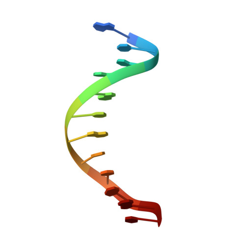
Binding of undamaged double stranded DNA to vaccinia virus uracil-DNA Glycosylase.

Schormann, N.Banerjee, S.Ricciardi, R.Chattopadhyay, D.

(2015) BMC Struct Biol 15: 10-10

  • DOI: https://doi.org/10.1186/s12900-015-0037-1
  • Primary Citation of Related Structures:  
    4QCB

  • PubMed Abstract: 

    Uracil-DNA glycosylases are evolutionarily conserved DNA repair enzymes. However, vaccinia virus uracil-DNA glycosylase (known as D4), also serves as an intrinsic and essential component of the processive DNA polymerase complex during DNA replication. In this complex D4 binds to a unique poxvirus specific protein A20 which tethers it to the DNA polymerase. At the replication fork the DNA scanning and repair function of D4 is coupled with DNA replication. So far, DNA-binding to D4 has not been structurally characterized. This manuscript describes the first structure of a DNA-complex of a uracil-DNA glycosylase from the poxvirus family. This also represents the first structure of a uracil DNA glycosylase in complex with an undamaged DNA. In the asymmetric unit two D4 subunits bind simultaneously to complementary strands of the DNA double helix. Each D4 subunit interacts mainly with the central region of one strand. DNA binds to the opposite side of the A20-binding surface on D4. Comparison of the present structure with the structure of uracil-containing DNA-bound human uracil-DNA glycosylase suggests that for DNA binding and uracil removal D4 employs a unique set of residues and motifs that are highly conserved within the poxvirus family but different in other organisms. The first structure of D4 bound to a truly non-specific undamaged double-stranded DNA suggests that initial binding of DNA may involve multiple non-specific interactions between the protein and the phosphate backbone.


  • Organizational Affiliation

    Department of Medicine, University of Alabama at Birmingham, Birmingham, AL, 35294, USA. nschorm@uab.edu.


Macromolecules

Find similar proteins by:  (by identity cutoff)  |  3D Structure
Entity ID: 1
MoleculeChains Sequence LengthOrganismDetailsImage
Uracil-DNA glycosylase
A, B
221Vaccinia virus WRMutation(s): 2 
Gene Names: VACWR109D4RUNG
EC: 3.2.2.27
UniProt
Find proteins for P04303 (Vaccinia virus (strain Western Reserve))
Explore P04303 
Go to UniProtKB:  P04303
Entity Groups  
Sequence Clusters30% Identity50% Identity70% Identity90% Identity95% Identity100% Identity
UniProt GroupP04303
Sequence Annotations
Expand
  • Reference Sequence

Find similar nucleic acids by:  Sequence   |   3D Structure  

Entity ID: 2
MoleculeChains LengthOrganismImage
5'-D(*GP*CP*AP*AP*AP*CP*GP*TP*TP*TP*GP*C)-3'
C, D
12N/A
Sequence Annotations
Expand
  • Reference Sequence
Experimental Data & Validation

Experimental Data

  • Method: X-RAY DIFFRACTION
  • Resolution: 2.89 Å
  • R-Value Free: 0.265 
  • R-Value Work: 0.216 
  • R-Value Observed: 0.218 
  • Space Group: P 21 21 21
Unit Cell:
Length ( Å )Angle ( ˚ )
a = 39.389α = 90
b = 92.327β = 90
c = 142.859γ = 90
Software Package:
Software NamePurpose
RAPDdata collection
PHASERphasing
REFMACrefinement
XDSdata reduction
SCALAdata scaling

Structure Validation

View Full Validation Report


Entry History 

Deposition Data

Revision History  (Full details and data files)

  • Version 1.0: 2015-06-10
    Type: Initial release
  • Version 1.1: 2016-06-29
    Changes: Source and taxonomy
  • Version 1.2: 2018-04-18
    Changes: Data collection, Database references
  • Version 1.3: 2023-09-20
    Changes: Data collection, Database references, Derived calculations, Refinement description