4OXD

Structure of the LdcB LD-carboxypeptidase reveals the molecular basis of peptidoglycan recognition


Experimental Data Snapshot

  • Method: X-RAY DIFFRACTION
  • Resolution: 2.80 Å
  • R-Value Free: 0.334 
  • R-Value Work: 0.273 
  • R-Value Observed: 0.276 

wwPDB Validation   3D Report Full Report


Ligand Structure Quality Assessment 


This is version 3.0 of the entry. See complete history


Literature

Structure of the LdcB LD-carboxypeptidase reveals the molecular basis of peptidoglycan recognition.

Hoyland, C.N.Aldridge, C.Cleverley, R.M.Duchene, M.C.Minasov, G.Onopriyenko, O.Sidiq, K.Stogios, P.J.Anderson, W.F.Daniel, R.A.Savchenko, A.Vollmer, W.Lewis, R.J.

(2014) Structure 22: 949-960

  • DOI: https://doi.org/10.1016/j.str.2014.04.015
  • Primary Citation of Related Structures:  
    4JID, 4MPH, 4OX3, 4OX5, 4OXD

  • PubMed Abstract: 

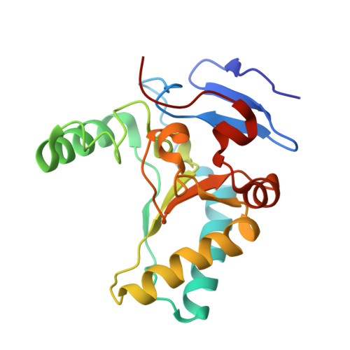
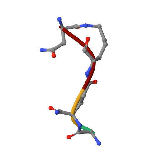
    Peptidoglycan surrounds the bacterial cytoplasmic membrane to protect the cell against osmolysis. The biosynthesis of peptidoglycan, made of glycan strands crosslinked by short peptides, is the target of antibiotics like β-lactams and glycopeptides. Nascent peptidoglycan contains pentapeptides that are trimmed by carboxypeptidases to tetra- and tripeptides. The well-characterized DD-carboxypeptidases hydrolyze the terminal D-alanine from the stem pentapeptide to produce a tetrapeptide. However, few LD-carboxypeptidases that produce tripeptides have been identified, and nothing is known about substrate specificity in these enzymes. We report biochemical properties and crystal structures of the LD-carboxypeptidases LdcB from Streptococcus pneumoniae, Bacillus anthracis, and Bacillus subtilis. The enzymes are active against bacterial cell wall tetrapeptides and adopt a zinc-carboxypeptidase fold characteristic of the LAS superfamily. We have also solved the structure of S. pneumoniae LdcB with a product mimic, elucidating the residues essential for peptidoglycan recognition and the conformational changes that occur on ligand binding.


  • Organizational Affiliation

    Institute for Cell and Molecular Biosciences, Newcastle University, Newcastle upon Tyne NE2 4HH, UK.


Macromolecules
Find similar proteins by:  (by identity cutoff)  |  3D Structure
Entity ID: 1
MoleculeChains Sequence LengthOrganismDetailsImage
LdcB LD-carboxypeptidase
A, B, C, D, E
187Streptococcus pneumoniae R6Mutation(s): 0 
Gene Names: spr0554
UniProt
Find proteins for Q8DQQ1 (Streptococcus pneumoniae (strain ATCC BAA-255 / R6))
Explore Q8DQQ1 
Go to UniProtKB:  Q8DQQ1
Entity Groups  
Sequence Clusters30% Identity50% Identity70% Identity90% Identity95% Identity100% Identity
UniProt GroupQ8DQQ1
Sequence Annotations
Expand
  • Reference Sequence

Find similar proteins by:  Sequence   |   3D Structure  

Entity ID: 2
MoleculeChains Sequence LengthOrganismDetailsImage
MUB-ALA-ZGL-LYS-DSGF [auth H]4Lactobacillus acidophilusMutation(s): 0 
Sequence Annotations
Expand
  • Reference Sequence
Small Molecules
Ligands 5 Unique
IDChains Name / Formula / InChI Key2D Diagram3D Interactions
MUB
Query on MUB

Download Ideal Coordinates CCD File 
CA [auth H]N-acetyl-alpha-muramic acid
C11 H19 N O8
MNLRQHMNZILYPY-MDMHTWEWSA-N
LYS
Query on LYS

Download Ideal Coordinates CCD File 
Q [auth B]LYSINE
C6 H15 N2 O2
KDXKERNSBIXSRK-YFKPBYRVSA-O
ZN
Query on ZN

Download Ideal Coordinates CCD File 
AA [auth E]
BA [auth E]
G [auth A]
H [auth A]
I [auth A]
AA [auth E],
BA [auth E],
G [auth A],
H [auth A],
I [auth A],
K [auth B],
L [auth B],
M [auth B],
N [auth B],
O [auth B],
P [auth B],
R [auth C],
S [auth C],
T [auth C],
U [auth C],
W [auth D],
X [auth D],
Z [auth E]
ZINC ION
Zn
PTFCDOFLOPIGGS-UHFFFAOYSA-N
CL
Query on CL

Download Ideal Coordinates CCD File 
V [auth C],
Y [auth D]
CHLORIDE ION
Cl
VEXZGXHMUGYJMC-UHFFFAOYSA-M
MG
Query on MG

Download Ideal Coordinates CCD File 
J [auth A]MAGNESIUM ION
Mg
JLVVSXFLKOJNIY-UHFFFAOYSA-N
Experimental Data & Validation

Experimental Data

  • Method: X-RAY DIFFRACTION
  • Resolution: 2.80 Å
  • R-Value Free: 0.334 
  • R-Value Work: 0.273 
  • R-Value Observed: 0.276 
  • Space Group: C 1 2 1
Unit Cell:
Length ( Å )Angle ( ˚ )
a = 345.954α = 90
b = 42.549β = 93.07
c = 79.318γ = 90
Software Package:
Software NamePurpose
REFMACrefinement

Structure Validation

View Full Validation Report


Ligand Structure Quality Assessment 


Entry History & Funding Information

Deposition Data


Funding OrganizationLocationGrant Number
Biotechnology and Biological Sciences Research Council (BBSRC)United Kingdom--

Revision History  (Full details and data files)

  • Version 1.0: 2014-05-21
    Type: Initial release
  • Version 1.1: 2014-07-16
    Changes: Database references
  • Version 1.2: 2014-10-01
    Changes: Database references
  • Version 1.3: 2015-01-07
    Changes: Database references
  • Version 1.4: 2020-01-01
    Changes: Author supporting evidence, Data collection, Database references, Derived calculations, Other, Source and taxonomy, Structure summary
  • Version 1.5: 2020-07-29
    Type: Remediation
    Reason: Carbohydrate remediation
    Changes: Data collection, Derived calculations, Refinement description, Structure summary
  • Version 2.0: 2023-11-15
    Changes: Atomic model, Data collection, Database references, Derived calculations, Structure summary
  • Version 3.0: 2024-04-10
    Changes: Atomic model, Data collection, Database references, Derived calculations, Polymer sequence, Source and taxonomy, Structure summary