4OL8

Ty3 reverse transcriptase bound to DNA/RNA


Experimental Data Snapshot

  • Method: X-RAY DIFFRACTION
  • Resolution: 3.10 Å
  • R-Value Free: 0.296 
  • R-Value Work: 0.227 
  • R-Value Observed: 0.231 

wwPDB Validation   3D Report Full Report


This is version 1.3 of the entry. See complete history


Literature

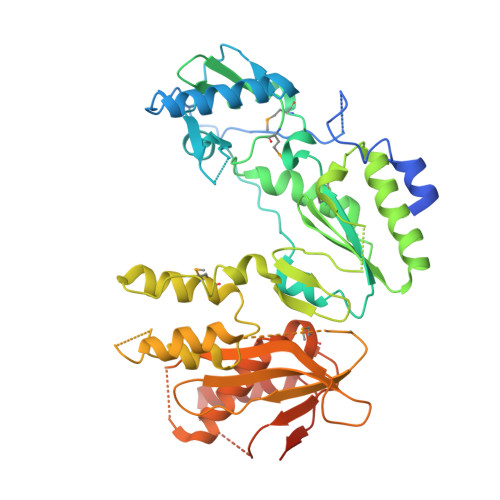
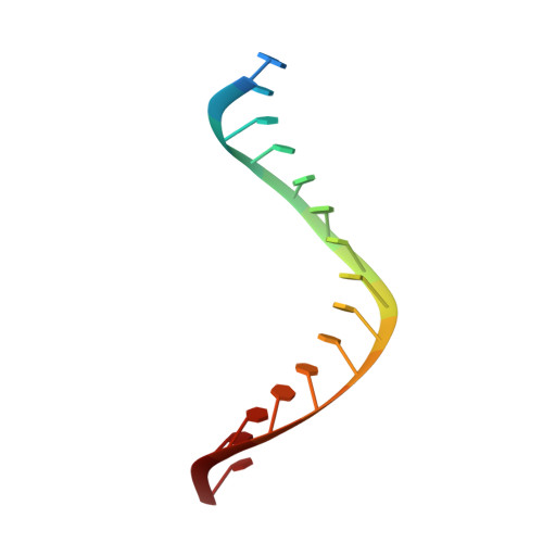
Ty3 reverse transcriptase complexed with an RNA-DNA hybrid shows structural and functional asymmetry.

Nowak, E.Miller, J.T.Bona, M.K.Studnicka, J.Szczepanowski, R.H.Jurkowski, J.Le Grice, S.F.Nowotny, M.

(2014) Nat Struct Mol Biol 21: 389-396

  • DOI: https://doi.org/10.1038/nsmb.2785
  • Primary Citation of Related Structures:  
    4OL8

  • PubMed Abstract: 

    Retrotransposons are a class of mobile genetic elements that replicate by converting their single-stranded RNA intermediate to double-stranded DNA through the combined DNA polymerase and ribonuclease H (RNase H) activities of the element-encoded reverse transcriptase (RT). Although a wealth of structural information is available for lentiviral and gammaretroviral RTs, equivalent studies on counterpart enzymes of long terminal repeat (LTR)-containing retrotransposons, from which they are evolutionarily derived, is lacking. In this study, we report the first crystal structure of a complex of RT from the Saccharomyces cerevisiae LTR retrotransposon Ty3 in the presence of its polypurine tract-containing RNA-DNA hybrid. In contrast to its retroviral counterparts, Ty3 RT adopts an asymmetric homodimeric architecture whose assembly is substrate dependent. Moreover, our structure and biochemical data suggest that the RNase H and DNA polymerase activities are contributed by individual subunits of the homodimer.


  • Organizational Affiliation

    Laboratory of Protein Structure, International Institute of Molecular and Cell Biology, Warsaw, Poland.


Macromolecules

Find similar proteins by:  (by identity cutoff)  |  3D Structure
Entity ID: 1
MoleculeChains Sequence LengthOrganismDetailsImage
Reverse transcriptase/ribonuclease HA,
B,
G [auth F],
H [auth E]
478Saccharomyces cerevisiaeMutation(s): 1 
Gene Names: TY3B-GYGRWTy3-1 POLYGR109W-BG5984
EC: 2.7.7.49 (PDB Primary Data), 2.7.7.7 (PDB Primary Data), 3.1.26.4 (PDB Primary Data)
UniProt
Find proteins for Q99315 (Saccharomyces cerevisiae (strain ATCC 204508 / S288c))
Explore Q99315 
Go to UniProtKB:  Q99315
Entity Groups  
Sequence Clusters30% Identity50% Identity70% Identity90% Identity95% Identity100% Identity
UniProt GroupQ99315
Sequence Annotations
Expand
  • Reference Sequence

Find similar nucleic acids by:  Sequence   |   3D Structure  

Entity ID: 2
MoleculeChains LengthOrganismImage
5'-R(*CP*UP*GP*AP*GP*AP*GP*AP*GP*AP*GP*GP*AP*AP*GP*AP*UP*G)-3'C,
E [auth G]
18N/A
Sequence Annotations
Expand
  • Reference Sequence

Find similar nucleic acids by:  Sequence   |   3D Structure  

Entity ID: 3
MoleculeChains LengthOrganismImage
5'-D(*CP*AP*TP*CP*TP*TP*CP*CP*TP*CP*TP*CP*TP*CP*TP*C)-3'D,
F [auth H]
16N/A
Sequence Annotations
Expand
  • Reference Sequence
Small Molecules
Modified Residues  1 Unique
IDChains TypeFormula2D DiagramParent
MSE
Query on MSE
A,
B,
G [auth F],
H [auth E]
L-PEPTIDE LINKINGC5 H11 N O2 SeMET
Experimental Data & Validation

Experimental Data

  • Method: X-RAY DIFFRACTION
  • Resolution: 3.10 Å
  • R-Value Free: 0.296 
  • R-Value Work: 0.227 
  • R-Value Observed: 0.231 
  • Space Group: P 21 21 2
Unit Cell:
Length ( Å )Angle ( ˚ )
a = 320.7α = 90
b = 75.062β = 90
c = 108.35γ = 90
Software Package:
Software NamePurpose
XDSdata scaling
AutoSolphasing
PHENIXrefinement
XDSdata reduction

Structure Validation

View Full Validation Report


Entry History 

Deposition Data

Revision History  (Full details and data files)

  • Version 1.0: 2014-03-05
    Type: Initial release
  • Version 1.1: 2014-03-12
    Changes: Structure summary
  • Version 1.2: 2014-04-02
    Changes: Database references
  • Version 1.3: 2014-04-16
    Changes: Database references