4NFY

Crystal Structure of 3-phosphoglycerate Dehydrogenase from Entamoeba histolytica


Experimental Data Snapshot

  • Method: X-RAY DIFFRACTION
  • Resolution: 2.45 Å
  • R-Value Free: 0.259 
  • R-Value Work: 0.194 
  • R-Value Observed: 0.197 

wwPDB Validation   3D Report Full Report


This is version 1.2 of the entry. See complete history


Literature

Crystal structures and kinetics of Type III 3-phosphoglycerate dehydrogenase reveal catalysis by lysine.

Singh, R.K.Raj, I.Pujari, R.Gourinath, S.

(2014) FEBS J 281: 5498-5512

  • DOI: https://doi.org/10.1111/febs.13091
  • Primary Citation of Related Structures:  
    4NFY, 4NJM, 4NJO

  • PubMed Abstract: 

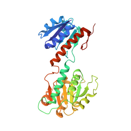
    D-Phosphoglycerate dehydrogenase (PGDH) catalyzes the first committed step of the phosphorylated serine biosynthesis pathway. Here, we report for the first time, the crystal structures of Type IIIK PGDH from Entamoeba histolytica in the apo form, as well as in complexes with substrate (3-phosphoglyceric acid) and cofactor (NAD(+) ) to 2.45, 1.8 and 2.2 Å resolution, respectively. Comparison of the apo structure with the substrate-bound structure shows that the substrate-binding domain is rotated by ~ 20° to close the active-site cleft. The cofactor-bound structure also shows a closed-cleft conformation, in which NAD(+) is bound to the nucleotide-binding domain and a formate ion occupies the substrate-binding site. Superposition of the substrate- and cofactor-bound structures represents a snapshot of the enzyme in the active form, where C2 of the substrate and C4N of the cofactor are 2.2 Å apart, and the amino group of Lys263 is close enough to the substrate to remove the proton from the hydroxyl group of PGA, indicating the role of Lys in the catalysis. Mutation of Lys263 to Ala yields just 0.8% of the specific activity of the wild-type enzyme, revealing that Lys263 indeed plays an integral role in the catalytic activity. The detectable activity of the mutant, however, indicates that after 20° rotation of the substrate-binding domain, the resulting positions of the substrate and cofactor are sufficiently close to make a productive reaction.


  • Organizational Affiliation

    School of Life Sciences, Jawaharlal Nehru University, New Delhi, India.


Macromolecules
Find similar proteins by:  (by identity cutoff)  |  3D Structure
Entity ID: 1
MoleculeChains Sequence LengthOrganismDetailsImage
D-3-phosphoglycerate dehydrogenase, putative
A, B
307Entamoeba histolyticaMutation(s): 0 
Gene Names: 3-phosphoglycerate dehydrogenaseEHI_060860EhPGDH
EC: 1.1.1.95
UniProt
Find proteins for Q76KF5 (Entamoeba histolytica)
Explore Q76KF5 
Go to UniProtKB:  Q76KF5
Entity Groups  
Sequence Clusters30% Identity50% Identity70% Identity90% Identity95% Identity100% Identity
UniProt GroupQ76KF5
Sequence Annotations
Expand
  • Reference Sequence
Small Molecules
Ligands 1 Unique
IDChains Name / Formula / InChI Key2D Diagram3D Interactions
TRS
Query on TRS

Download Ideal Coordinates CCD File 
C [auth A],
D [auth B]
2-AMINO-2-HYDROXYMETHYL-PROPANE-1,3-DIOL
C4 H12 N O3
LENZDBCJOHFCAS-UHFFFAOYSA-O
Experimental Data & Validation

Experimental Data

  • Method: X-RAY DIFFRACTION
  • Resolution: 2.45 Å
  • R-Value Free: 0.259 
  • R-Value Work: 0.194 
  • R-Value Observed: 0.197 
  • Space Group: P 41
Unit Cell:
Length ( Å )Angle ( ˚ )
a = 85.906α = 90
b = 85.906β = 90
c = 84.492γ = 90
Software Package:
Software NamePurpose
HKL-2000data collection
MOLREPphasing
REFMACrefinement
HKL-2000data reduction
SCALEPACKdata scaling

Structure Validation

View Full Validation Report


Entry History 

Deposition Data

Revision History  (Full details and data files)

  • Version 1.0: 2014-10-08
    Type: Initial release
  • Version 1.1: 2015-06-17
    Changes: Database references
  • Version 1.2: 2023-11-08
    Changes: Data collection, Database references, Derived calculations, Refinement description