4LSI

Ion selectivity of OmpF porin soaked in 0.3M KBr


Experimental Data Snapshot

  • Method: X-RAY DIFFRACTION
  • Resolution: 2.09 Å
  • R-Value Free: 0.245 
  • R-Value Work: 0.190 
  • R-Value Observed: 0.193 

wwPDB Validation   3D Report Full Report


Ligand Structure Quality Assessment 


This is version 1.2 of the entry. See complete history


Literature

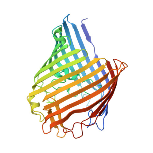
A structural study of ion permeation in OmpF porin from anomalous X-ray diffraction and molecular dynamics simulations.

Dhakshnamoorthy, B.Ziervogel, B.K.Blachowicz, L.Roux, B.

(2013) J Am Chem Soc 135: 16561-16568

  • DOI: https://doi.org/10.1021/ja407783a
  • Primary Citation of Related Structures:  
    4LSE, 4LSF, 4LSH, 4LSI

  • PubMed Abstract: 

    OmpF, a multiionic porin from Escherichia coli, is a useful protypical model system for addressing general questions about electrostatic interactions in the confinement of an aqueous molecular pore. Here, favorable anion locations in the OmpF pore were mapped by anomalous X-ray scattering of Br(–) ions from four different crystal structures and compared with Mg(2+) sites and Rb(+) sites from a previous anomalous diffraction study to provide a complete picture of cation and anion transfer paths along the OmpF channel. By comparing structures with various crystallization conditions, we find that anions bind in discrete clusters along the entire length of the OmpF pore, whereas cations find conserved binding sites with the extracellular, surface-exposed loops. Results from molecular dynamics simulations are consistent with the experimental data and help highlight the critical residues that preferentially contact either cations or anions during permeation. Analysis of these results provides new insights into the molecular mechanisms that determine ion selectivity in OmpF porin.


  • Organizational Affiliation

    Department of Biochemistry and Molecular Biology, Gordon Center for Integrative Science, University of Chicago Chicago, IL 60637, USA.


Macromolecules
Find similar proteins by:  (by identity cutoff)  |  3D Structure
Entity ID: 1
MoleculeChains Sequence LengthOrganismDetailsImage
Outer membrane protein F
A, B, C
341Escherichia coli K-12Mutation(s): 0 
Gene Names: b0929cmlBcoacryJW0912ompFtolF
Membrane Entity: Yes 
UniProt
Find proteins for P02931 (Escherichia coli (strain K12))
Explore P02931 
Go to UniProtKB:  P02931
Entity Groups  
Sequence Clusters30% Identity50% Identity70% Identity90% Identity95% Identity100% Identity
UniProt GroupP02931
Sequence Annotations
Expand
  • Reference Sequence
Small Molecules
Ligands 5 Unique
IDChains Name / Formula / InChI Key2D Diagram3D Interactions
C8E
Query on C8E

Download Ideal Coordinates CCD File 
AA [auth C],
F [auth A],
S [auth B],
T [auth B]
(HYDROXYETHYLOXY)TRI(ETHYLOXY)OCTANE
C16 H34 O5
FEOZZFHAVXYAMB-UHFFFAOYSA-N
PEG
Query on PEG

Download Ideal Coordinates CCD File 
D [auth A]
E [auth A]
FA [auth C]
GA [auth C]
HA [auth C]
D [auth A],
E [auth A],
FA [auth C],
GA [auth C],
HA [auth C],
O [auth A],
P [auth B],
Q [auth B],
R [auth B]
DI(HYDROXYETHYL)ETHER
C4 H10 O3
MTHSVFCYNBDYFN-UHFFFAOYSA-N
GOL
Query on GOL

Download Ideal Coordinates CCD File 
G [auth A],
H [auth A],
IA [auth C],
U [auth B]
GLYCEROL
C3 H8 O3
PEDCQBHIVMGVHV-UHFFFAOYSA-N
BR
Query on BR

Download Ideal Coordinates CCD File 
JA [auth C]
K [auth A]
KA [auth C]
L [auth A]
M [auth A]
JA [auth C],
K [auth A],
KA [auth C],
L [auth A],
M [auth A],
N [auth A],
X [auth B],
Y [auth B],
Z [auth B]
BROMIDE ION
Br
CPELXLSAUQHCOX-UHFFFAOYSA-M
MG
Query on MG

Download Ideal Coordinates CCD File 
BA [auth C]
CA [auth C]
DA [auth C]
EA [auth C]
I [auth A]
BA [auth C],
CA [auth C],
DA [auth C],
EA [auth C],
I [auth A],
J [auth A],
V [auth B],
W [auth B]
MAGNESIUM ION
Mg
JLVVSXFLKOJNIY-UHFFFAOYSA-N
Experimental Data & Validation

Experimental Data

  • Method: X-RAY DIFFRACTION
  • Resolution: 2.09 Å
  • R-Value Free: 0.245 
  • R-Value Work: 0.190 
  • R-Value Observed: 0.193 
  • Space Group: C 1 2 1
Unit Cell:
Length ( Å )Angle ( ˚ )
a = 202.094α = 90
b = 116.559β = 97
c = 53.592γ = 90
Software Package:
Software NamePurpose
ADSCdata collection
PHASERphasing
REFMACrefinement
HKL-2000data reduction
HKL-2000data scaling

Structure Validation

View Full Validation Report


Ligand Structure Quality Assessment 


Entry History 

Deposition Data

Revision History  (Full details and data files)

  • Version 1.0: 2013-10-23
    Type: Initial release
  • Version 1.1: 2014-01-15
    Changes: Database references
  • Version 1.2: 2023-09-20
    Changes: Data collection, Database references, Derived calculations, Refinement description