4LL9

Crystal structure of D3D4 domain of the LILRB1 molecule


Experimental Data Snapshot

  • Method: X-RAY DIFFRACTION
  • Resolution: 2.69 Å
  • R-Value Free: 0.275 
  • R-Value Work: 0.245 
  • R-Value Observed: 0.246 

wwPDB Validation   3D Report Full Report


This is version 1.1 of the entry. See complete history


Literature

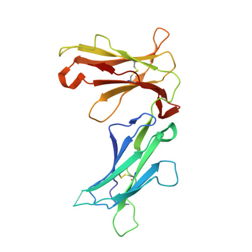
Crystal structures of the two membrane-proximal Ig-like domains (D3D4) of LILRB1/B2: alternative models for their involvement in peptide-HLA binding

Nam, G.Shi, Y.Ryu, M.Wang, Q.Song, H.Liu, J.Yan, J.Qi, J.Gao, G.F.

(2013) Protein Cell 4: 761-770

  • DOI: https://doi.org/10.1007/s13238-013-3908-x
  • Primary Citation of Related Structures:  
    4LL9, 4LLA

  • PubMed Abstract: 

    Leukocyte immunoglobulin-like receptors (LILRs), also called CD85s, ILTs, or LIRs, are important mediators of immune activation and tolerance that contain tandem immunoglobulin (Ig)-like folds. There are 11 (in addition to two pseudogenes) LILRs in total, two with two Ig-like domains (D1D2) and the remaining nine with four Ig-like domains (D1D2D3D4). Thus far, the structural features of the D1D2 domains of LILR proteins are well defined, but no structures for the D3D4 domains have been reported. This is a very important field to be studied as it relates to the unknown functions of the D3D4 domains, as well as their relative orientation to the D1D2 domains on the cell surface. Here, we report the crystal structures of the D3D4 domains of both LILRB1 and LILRB2. The two Ig-like domains of both LILRB1-D3D4 and LILRB2-D3D4 are arranged at an acute angle (∼60°) to form a bent structure, resembling the structures of natural killer inhibitory receptors. Based on these two D3D4 domain structures and previously reported D1D2/HLA I complex structures, two alternative models of full-length (four Ig-like domains) LILR molecules bound to HLA I are proposed.


  • Organizational Affiliation

    CAS Key Laboratory of Pathogenic Microbiology and Immunology, Institute of Microbiology, Chinese Academy of Sciences, Beijing, 100101, China.


Macromolecules
Find similar proteins by:  (by identity cutoff)  |  3D Structure
Entity ID: 1
MoleculeChains Sequence LengthOrganismDetailsImage
Leukocyte immunoglobulin-like receptor subfamily B member 1
A, B, C
196Homo sapiensMutation(s): 0 
Gene Names: LILRB1
UniProt & NIH Common Fund Data Resources
Find proteins for Q8NHL6 (Homo sapiens)
Explore Q8NHL6 
Go to UniProtKB:  Q8NHL6
PHAROS:  Q8NHL6
Entity Groups  
Sequence Clusters30% Identity50% Identity70% Identity90% Identity95% Identity100% Identity
UniProt GroupQ8NHL6
Sequence Annotations
Expand
  • Reference Sequence
Small Molecules
Ligands 1 Unique
IDChains Name / Formula / InChI Key2D Diagram3D Interactions
IOD
Query on IOD

Download Ideal Coordinates CCD File 
D [auth A]
E [auth A]
F [auth A]
G [auth A]
H [auth A]
D [auth A],
E [auth A],
F [auth A],
G [auth A],
H [auth A],
I [auth B],
J [auth B],
K [auth B],
L [auth B],
M [auth C],
N [auth C],
O [auth C]
IODIDE ION
I
XMBWDFGMSWQBCA-UHFFFAOYSA-M
Experimental Data & Validation

Experimental Data

  • Method: X-RAY DIFFRACTION
  • Resolution: 2.69 Å
  • R-Value Free: 0.275 
  • R-Value Work: 0.245 
  • R-Value Observed: 0.246 
  • Space Group: C 1 2 1
Unit Cell:
Length ( Å )Angle ( ˚ )
a = 121.257α = 90
b = 71.976β = 95.2
c = 75.036γ = 90
Software Package:
Software NamePurpose
ADSCdata collection
SHELXSphasing
PHENIXrefinement
HKL-2000data reduction
HKL-2000data scaling

Structure Validation

View Full Validation Report


Entry History 

Deposition Data

Revision History  (Full details and data files)

  • Version 1.0: 2013-09-11
    Type: Initial release
  • Version 1.1: 2013-11-06
    Changes: Database references