4LIZ

Crystal structure of coactosin from Entamoeba histolytica


Experimental Data Snapshot

  • Method: X-RAY DIFFRACTION
  • Resolution: 1.50 Å
  • R-Value Free: 0.188 
  • R-Value Work: 0.162 
  • R-Value Observed: 0.163 

wwPDB Validation   3D Report Full Report


This is version 1.2 of the entry. See complete history


Literature

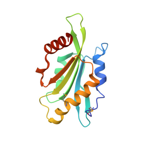
EhCoactosin stabilizes actin filaments in the protist parasite Entamoeba histolytica.

Kumar, N.SomlataMazumder, M.Dutta, P.Maiti, S.Gourinath, S.

(2014) PLoS Pathog 10: e1004362-e1004362

  • DOI: https://doi.org/10.1371/journal.ppat.1004362
  • Primary Citation of Related Structures:  
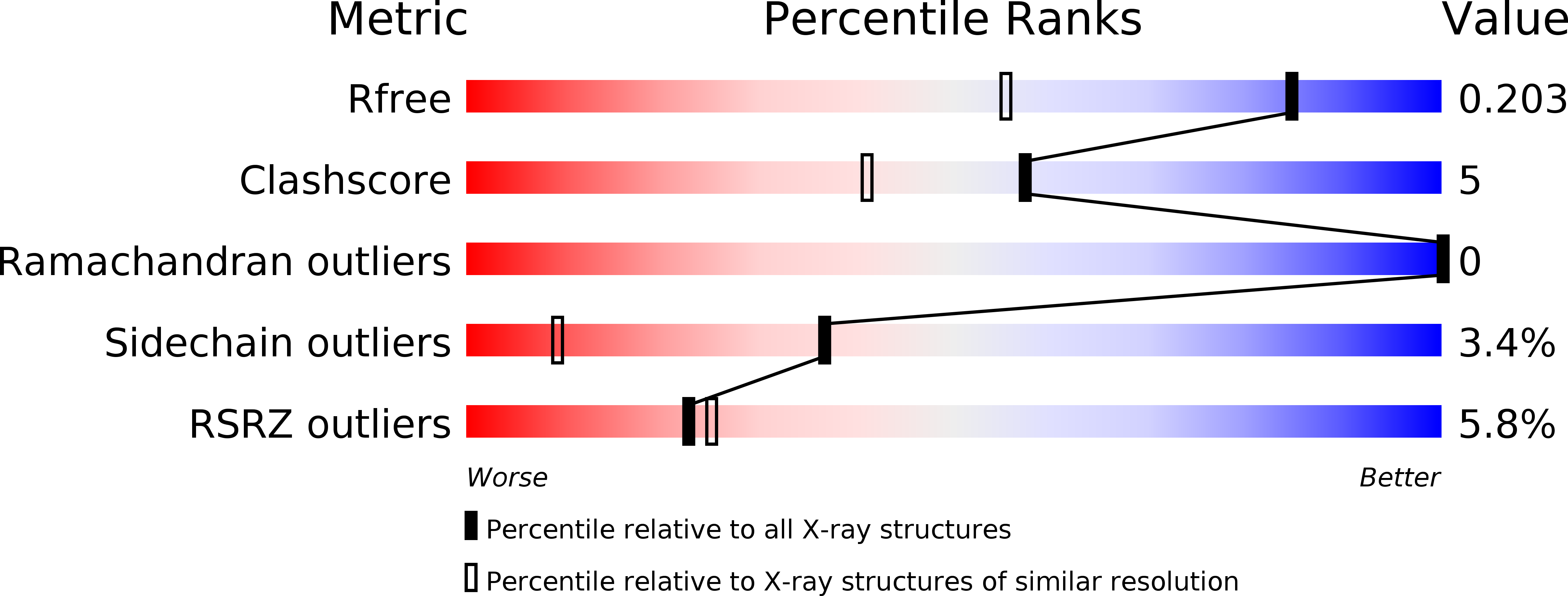
    4LIZ

  • PubMed Abstract: 

    Entamoeba histolytica is a protist parasite that is the causative agent of amoebiasis, and is a highly motile organism. The motility is essential for its survival and pathogenesis, and a dynamic actin cytoskeleton is required for this process. EhCoactosin, an actin-binding protein of the ADF/cofilin family, participates in actin dynamics, and here we report our studies of this protein using both structural and functional approaches. The X-ray crystal structure of EhCoactosin resembles that of human coactosin-like protein, with major differences in the distribution of surface charges and the orientation of terminal regions. According to in vitro binding assays, full-length EhCoactosin binds both F- and G-actin. Instead of acting to depolymerize or severe F-actin, EhCoactosin directly stabilizes the polymer. When EhCoactosin was visualized in E. histolytica cells using either confocal imaging or total internal reflectance microscopy, it was found to colocalize with F-actin at phagocytic cups. Over-expression of this protein stabilized F-actin and inhibited the phagocytic process. EhCoactosin appears to be an unusual type of coactosin involved in E. histolytica actin dynamics.


  • Organizational Affiliation

    School of Life Sciences, Jawaharlal Nehru University, New Delhi, India.


Macromolecules
Find similar proteins by:  (by identity cutoff)  |  3D Structure
Entity ID: 1
MoleculeChains Sequence LengthOrganismDetailsImage
Actin-binding protein, cofilin/tropomyosin family protein, putative142Entamoeba histolytica HM-1:IMSS-AMutation(s): 0 
Gene Names: Actin binding proteinCofilin/tropomyosin familyEHI7A_006660
UniProt
Find proteins for C4M4P4 (Entamoeba histolytica (strain ATCC 30459 / HM-1:IMSS / ABRM))
Explore C4M4P4 
Go to UniProtKB:  C4M4P4
Entity Groups  
Sequence Clusters30% Identity50% Identity70% Identity90% Identity95% Identity100% Identity
UniProt GroupC4M4P4
Sequence Annotations
Expand
  • Reference Sequence
Small Molecules
Ligands 2 Unique
IDChains Name / Formula / InChI Key2D Diagram3D Interactions
EPE
Query on EPE

Download Ideal Coordinates CCD File 
B [auth A]4-(2-HYDROXYETHYL)-1-PIPERAZINE ETHANESULFONIC ACID
C8 H18 N2 O4 S
JKMHFZQWWAIEOD-UHFFFAOYSA-N
NA
Query on NA

Download Ideal Coordinates CCD File 
C [auth A]SODIUM ION
Na
FKNQFGJONOIPTF-UHFFFAOYSA-N
Modified Residues  1 Unique
IDChains TypeFormula2D DiagramParent
MSE
Query on MSE
A
L-PEPTIDE LINKINGC5 H11 N O2 SeMET
Experimental Data & Validation

Experimental Data

  • Method: X-RAY DIFFRACTION
  • Resolution: 1.50 Å
  • R-Value Free: 0.188 
  • R-Value Work: 0.162 
  • R-Value Observed: 0.163 
  • Space Group: P 65
Unit Cell:
Length ( Å )Angle ( ˚ )
a = 76.601α = 90
b = 76.601β = 90
c = 54.647γ = 120
Software Package:
Software NamePurpose
DENZOdata reduction
SCALEPACKdata scaling
SHELXphasing
REFMACrefinement
PDB_EXTRACTdata extraction
HKL-2000data collection
SHELXDphasing

Structure Validation

View Full Validation Report


Entry History 

Deposition Data

Revision History  (Full details and data files)

  • Version 1.0: 2014-07-23
    Type: Initial release
  • Version 1.1: 2015-02-11
    Changes: Database references
  • Version 1.2: 2017-11-15
    Changes: Refinement description