4IL1

Crystal Structure of the Rat Calcineurin


Experimental Data Snapshot

  • Method: X-RAY DIFFRACTION
  • Resolution: 3.00 Å
  • R-Value Free: 0.273 
  • R-Value Work: 0.241 
  • R-Value Observed: 0.242 

wwPDB Validation   3D Report Full Report


This is version 1.3 of the entry. See complete history


Literature

Structural basis of calcineurin activation by calmodulin.

Ye, Q.Feng, Y.Yin, Y.Faucher, F.Currie, M.A.Rahman, M.N.Jin, J.Li, S.Wei, Q.Jia, Z.

(2013) Cell Signal 25: 2661-2667

  • DOI: https://doi.org/10.1016/j.cellsig.2013.08.033
  • Primary Citation of Related Structures:  
    4IL1

  • PubMed Abstract: 

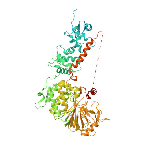
    Calcineurin is the only known calmodulin (CaM) activated protein phosphatase, which is involved in the regulation of numerous cellular and developmental processes and in calcium-dependent signal transduction. Although commonly assumed that CaM displaces the autoinhibitory domain (AID) blocking substrate access to its active site, the structural basis underlying activation remains elusive. We have created a fused ternary complex (CBA) by covalently linking three polypeptides: CaM, calcineurin regulatory B subunit (CnB) and calcineurin catalytic A subunit (CnA). CBA catalytic activity is comparable to that of fully activated native calcineurin in the presence of CaM. The crystal structure showed virtually no structural change in the active site and no evidence of CaM despite being covalently linked. The asymmetric unit contains four molecules; two parallel CBA pairs are packed in an antiparallel mode and the large cavities in crystal packing near the calcineurin active site would easily accommodate multiple positions of AID-bound CaM. Intriguingly, the conformation of the ordered segment of AID is not altered by CaM; thus, it is the disordered part of AID, which resumes a regular α-helical conformation upon binding to CaM, which is displaced by CaM for activation. We propose that the structural basis of calcineurin activation by CaM is through displacement of the disordered fragment of AID which otherwise impedes active site access.


  • Organizational Affiliation

    College of Chemistry, Beijing Normal University, Beijing 100875, People's Republic of China; Department of Biomedical and Molecular Sciences, Queen's University, 18 Stuart Street, Kingston, Ontario K7L 3N6, Canada.


Macromolecules
Find similar proteins by:  (by identity cutoff)  |  3D Structure
Entity ID: 1
MoleculeChains Sequence LengthOrganismDetailsImage
Calmodulin, Calcineurin subunit B type 1, Serine/threonine-protein phosphatase 2B catalytic subunit alpha isoform
A, B, C, D
823Rattus norvegicusMutation(s): 0 
Gene Names: 
CalmCalm1Calm2Calm3CalnaCamCam1Cam2Cam3Camb...
CalmCalm1Calm2Calm3CalnaCamCam1Cam2Cam3CambCamcPpp3caCn a2CnbPpp3r1

EC: 3.1.3.16
UniProt
Find proteins for P63100 (Rattus norvegicus)
Explore P63100 
Go to UniProtKB:  P63100
Find proteins for P63329 (Rattus norvegicus)
Explore P63329 
Go to UniProtKB:  P63329
Find proteins for P0DP29 (Rattus norvegicus)
Explore P0DP29 
Go to UniProtKB:  P0DP29
Entity Groups  
Sequence Clusters30% Identity50% Identity70% Identity90% Identity95% Identity100% Identity
UniProt GroupsP63329P0DP29P63100
Sequence Annotations
Expand
  • Reference Sequence
Small Molecules
Ligands 1 Unique
IDChains Name / Formula / InChI Key2D Diagram3D Interactions
CA
Query on CA

Download Ideal Coordinates CCD File 
E [auth A]
F [auth A]
G [auth A]
H [auth A]
I [auth B]
E [auth A],
F [auth A],
G [auth A],
H [auth A],
I [auth B],
J [auth B],
K [auth B],
L [auth B],
M [auth C],
N [auth C],
O [auth C],
P [auth C],
Q [auth D],
R [auth D],
S [auth D],
T [auth D]
CALCIUM ION
Ca
BHPQYMZQTOCNFJ-UHFFFAOYSA-N
Experimental Data & Validation

Experimental Data

  • Method: X-RAY DIFFRACTION
  • Resolution: 3.00 Å
  • R-Value Free: 0.273 
  • R-Value Work: 0.241 
  • R-Value Observed: 0.242 
  • Space Group: P 1 21 1
Unit Cell:
Length ( Å )Angle ( ˚ )
a = 99.3α = 90
b = 193.85β = 90.09
c = 107.82γ = 90
Software Package:
Software NamePurpose
ADSCdata collection
PHASESphasing
REFMACrefinement
XDSdata reduction
SCALAdata scaling

Structure Validation

View Full Validation Report


Entry History 

Deposition Data

Revision History  (Full details and data files)

  • Version 1.0: 2013-10-02
    Type: Initial release
  • Version 1.1: 2013-10-09
    Changes: Database references
  • Version 1.2: 2017-08-09
    Changes: Refinement description, Source and taxonomy
  • Version 1.3: 2023-09-20
    Changes: Data collection, Database references, Derived calculations, Refinement description