4HLY

The complex crystal structure of the DNA binding domain of vIRF-1 from the oncogenic KSHV with DNA


Experimental Data Snapshot

  • Method: X-RAY DIFFRACTION
  • Resolution: 1.48 Å
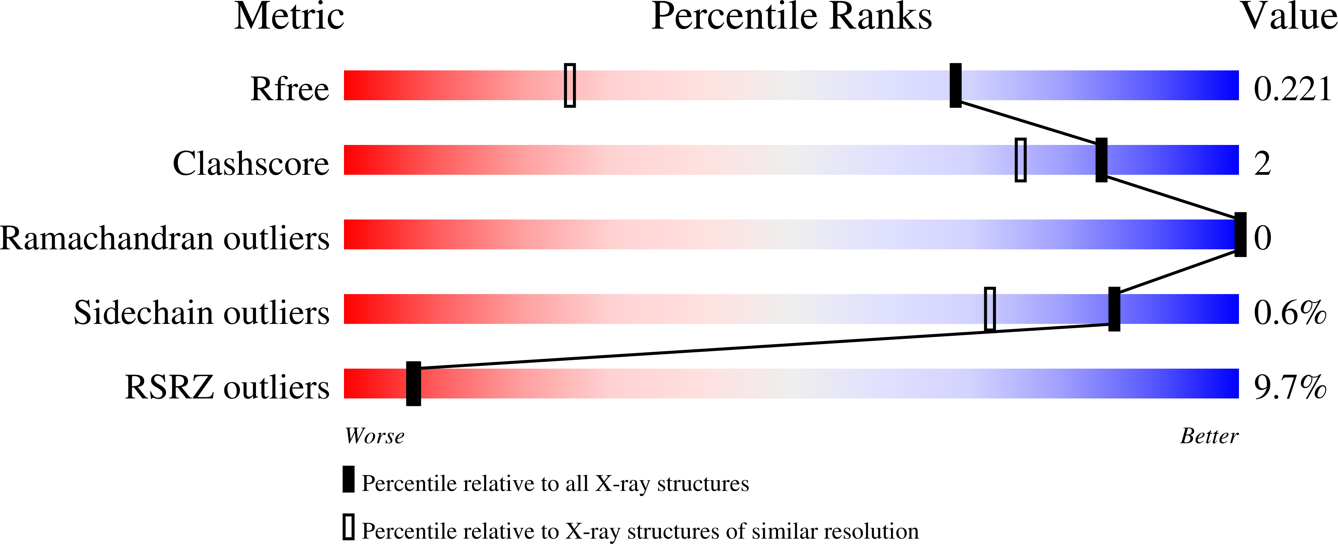
  • R-Value Free: 0.228 
  • R-Value Work: 0.196 
  • R-Value Observed: 0.198 

wwPDB Validation   3D Report Full Report


This is version 1.4 of the entry. See complete history


Literature

The crystal structure of the DNA-binding domain of vIRF-1 from the oncogenic KSHV reveals a conserved fold for DNA binding and reinforces its role as a transcription factor.

Hew, K.Dahlroth, S.L.Venkatachalam, R.Nasertorabi, F.Lim, B.T.Cornvik, T.Nordlund, P.

(2013) Nucleic Acids Res 41: 4295-4306

  • DOI: https://doi.org/10.1093/nar/gkt082
  • Primary Citation of Related Structures:  
    4HLX, 4HLY

  • PubMed Abstract: 

    Kaposi's sarcoma-associated herpesvirus encodes four viral homologues to cellular interferon regulatory factors (IRFs), where the most studied is vIRF-1. Even though vIRF-1 shows sequence homology to the N-terminal DNA-binding domain (DBD) of human IRFs, a specific role for this domain in vIRF-1's function has remained uncertain. To provide insights into the function of the vIRF-1 DBD, we have determined the crystal structure of it in complex with DNA and in its apo-form. Using a thermal stability shift assay (TSSA), we show that the vIRF-1 DBD binds DNA, whereas full-length vIRF-1 does not, suggesting a cis-acting regulatory mechanism in similarity to human IRFs. The complex structure of vIRF-1 DBD reveals interactions with the DNA backbone and the positioning of two arginines for specific recognition in the major grove. A superimposition with human IRF-3 reveals a similar positioning of the two specificity-determining arginines, and additional TSSAs indicate binding of vIRF-1 to an IRF-3 operator consensus sequence. The results from this study, therefore, provide support that vIRF-1 has evolved to bind DNA and plays a role in DNA binding in the context of transcriptional regulation and might act on some of the many operator sequences controlled by human IRF-3.


  • Organizational Affiliation

    Division of Structural Biology and Biochemistry, School of Biological Sciences, Nanyang Technological University, 138673 Singapore, Singapore.


Macromolecules

Find similar proteins by:  (by identity cutoff)  |  3D Structure
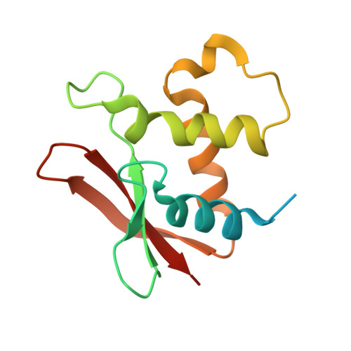
Entity ID: 1
MoleculeChains Sequence LengthOrganismDetailsImage
K9
A, B
132Human gammaherpesvirus 8Mutation(s): 0 
Gene Names: ORF K9vIRFvIRF-1
UniProt
Find proteins for F5HF68 (Human herpesvirus 8 type P (isolate GK18))
Explore F5HF68 
Go to UniProtKB:  F5HF68
Entity Groups  
Sequence Clusters30% Identity50% Identity70% Identity90% Identity95% Identity100% Identity
UniProt GroupF5HF68
Sequence Annotations
Expand
  • Reference Sequence

Find similar nucleic acids by:  Sequence   |   3D Structure  

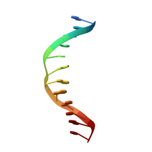
Entity ID: 2
MoleculeChains LengthOrganismImage
5'-D(*GP*CP*GP*TP*CP*GP*AP*GP*AP*CP*GP*C)-3'
C, D
12synthetic construct
Sequence Annotations
Expand
  • Reference Sequence
Experimental Data & Validation

Experimental Data

  • Method: X-RAY DIFFRACTION
  • Resolution: 1.48 Å
  • R-Value Free: 0.228 
  • R-Value Work: 0.196 
  • R-Value Observed: 0.198 
  • Space Group: P 1
Unit Cell:
Length ( Å )Angle ( ˚ )
a = 38.536α = 91.32
b = 38.577β = 88.48
c = 48.562γ = 63.13
Software Package:
Software NamePurpose
HKL-2000data collection
PHASERphasing
REFMACrefinement
HKL-2000data reduction
HKL-2000data scaling

Structure Validation

View Full Validation Report


Entry History 

Deposition Data

Revision History  (Full details and data files)

  • Version 1.0: 2013-03-13
    Type: Initial release
  • Version 1.1: 2014-02-05
    Changes: Database references
  • Version 1.2: 2016-09-07
    Changes: Derived calculations, Source and taxonomy
  • Version 1.3: 2016-09-14
    Changes: Derived calculations
  • Version 1.4: 2024-03-20
    Changes: Data collection, Database references