4GP4

Structure of Recombinant Cytochrome ba3 Oxidase mutant Y133F from Thermus thermophilus


Experimental Data Snapshot

  • Method: X-RAY DIFFRACTION
  • Resolution: 2.80 Å
  • R-Value Free: 0.227 
  • R-Value Work: 0.179 
  • R-Value Observed: 0.182 

wwPDB Validation   3D Report Full Report


Ligand Structure Quality Assessment 


This is version 1.4 of the entry. See complete history


Literature

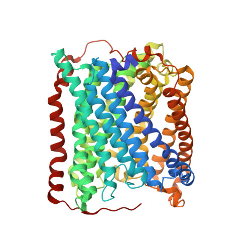
Ligand Access to the Active Site in Thermus thermophilusba(3) and Bovine Heart aa(3) Cytochrome Oxidases.

McDonald, W.Funatogawa, C.Li, Y.Szundi, I.Chen, Y.Fee, J.A.Stout, C.D.Einarsdottir, O.

(2013) Biochemistry 52: 640-652

  • DOI: https://doi.org/10.1021/bi301358a
  • Primary Citation of Related Structures:  
    4GP4, 4GP5, 4GP8

  • PubMed Abstract: 

    Knowledge of the structure and dynamics of the ligand channel(s) in heme-copper oxidases is critical for understanding how the protein environment modulates the functions of these enzymes. Using photolabile NO and O(2) carriers, we recently found that NO and O(2) binding in Thermus thermophilus (Tt) ba(3) is ~10 times faster than in the bovine enzyme, indicating that inherent structural differences affect ligand access in these enzymes. Using X-ray crystallography, time-resolved optical absorption measurements, and theoretical calculations, we investigated ligand access in wild-type Tt ba(3) and the mutants, Y133W, T231F, and Y133W/T231F, in which tyrosine and threonine in the O(2) channel of Tt ba(3) are replaced by the corresponding bulkier tryptophan and phenylalanine, respectively, present in the aa(3) enzymes. NO binding in Y133W and Y133W/T231F was found to be 5 times slower than in wild-type ba(3) and the T231F mutant. The results show that the Tt ba(3) Y133W mutation and the bovine W126 residue physically impede NO access to the binuclear center. In the bovine enzyme, there is a hydrophobic "way station", which may further slow ligand access to the active site. Classical simulations of diffusion of Xe to the active sites in ba(3) and bovine aa(3) show conformational freedom of the bovine F238 and the F231 side chain of the Tt ba(3) Y133W/T231F mutant, with both residues rotating out of the ligand channel, resulting in no effect on ligand access in either enzyme.


  • Organizational Affiliation

    Department of Chemistry and Biochemistry, University of California, Santa Cruz, CA 95064, USA.


Macromolecules
Find similar proteins by:  (by identity cutoff)  |  3D Structure
Entity ID: 1
MoleculeChains Sequence LengthOrganismDetailsImage
Cytochrome c oxidase subunit 1568Thermus thermophilus HB8Mutation(s): 1 
Gene Names: cbaATTHA1135
EC: 1.9.3.1
Membrane Entity: Yes 
UniProt
Find proteins for Q5SJ79 (Thermus thermophilus (strain ATCC 27634 / DSM 579 / HB8))
Explore Q5SJ79 
Go to UniProtKB:  Q5SJ79
Entity Groups  
Sequence Clusters30% Identity50% Identity70% Identity90% Identity95% Identity100% Identity
UniProt GroupQ5SJ79
Sequence Annotations
Expand
  • Reference Sequence
Find similar proteins by:  (by identity cutoff)  |  3D Structure
Entity ID: 2
MoleculeChains Sequence LengthOrganismDetailsImage
Cytochrome c oxidase subunit 2168Thermus thermophilus HB8Mutation(s): 0 
Gene Names: cbaBctaCTTHA1134
EC: 1.9.3.1
Membrane Entity: Yes 
UniProt
Find proteins for Q5SJ80 (Thermus thermophilus (strain ATCC 27634 / DSM 579 / HB8))
Explore Q5SJ80 
Go to UniProtKB:  Q5SJ80
Entity Groups  
Sequence Clusters30% Identity50% Identity70% Identity90% Identity95% Identity100% Identity
UniProt GroupQ5SJ80
Sequence Annotations
Expand
  • Reference Sequence
Find similar proteins by:  (by identity cutoff)  |  3D Structure
Entity ID: 3
MoleculeChains Sequence LengthOrganismDetailsImage
Cytochrome c oxidase polypeptide 2A34Thermus thermophilus HB8Mutation(s): 0 
Gene Names: cbaDTTHA1133
EC: 1.9.3.1
Membrane Entity: Yes 
UniProt
Find proteins for P82543 (Thermus thermophilus (strain ATCC 27634 / DSM 579 / HB8))
Explore P82543 
Go to UniProtKB:  P82543
Entity Groups  
Sequence Clusters30% Identity50% Identity70% Identity90% Identity95% Identity100% Identity
UniProt GroupP82543
Sequence Annotations
Expand
  • Reference Sequence
Small Molecules
Ligands 6 Unique
IDChains Name / Formula / InChI Key2D Diagram3D Interactions
HAS
Query on HAS

Download Ideal Coordinates CCD File 
F [auth A]HEME-AS
C54 H64 Fe N4 O6
PDYODZVCODUKFH-ZOMLSHGTSA-L
HEM
Query on HEM

Download Ideal Coordinates CCD File 
E [auth A]PROTOPORPHYRIN IX CONTAINING FE
C34 H32 Fe N4 O4
KABFMIBPWCXCRK-RGGAHWMASA-L
OLC
Query on OLC

Download Ideal Coordinates CCD File 
H [auth A]
I [auth A]
J [auth A]
K [auth A]
L [auth A]
H [auth A],
I [auth A],
J [auth A],
K [auth A],
L [auth A],
M [auth A],
N [auth A],
O [auth A],
P [auth A],
Q [auth A],
R [auth A],
S [auth A],
T [auth A],
V [auth B],
W [auth B],
X [auth B]
(2R)-2,3-dihydroxypropyl (9Z)-octadec-9-enoate
C21 H40 O4
RZRNAYUHWVFMIP-GDCKJWNLSA-N
CUA
Query on CUA

Download Ideal Coordinates CCD File 
U [auth B]DINUCLEAR COPPER ION
Cu2
ALKZAGKDWUSJED-UHFFFAOYSA-N
CU
Query on CU

Download Ideal Coordinates CCD File 
D [auth A]COPPER (II) ION
Cu
JPVYNHNXODAKFH-UHFFFAOYSA-N
PER
Query on PER

Download Ideal Coordinates CCD File 
G [auth A]PEROXIDE ION
O2
ANAIPYUSIMHBEL-UHFFFAOYSA-N
Experimental Data & Validation

Experimental Data

  • Method: X-RAY DIFFRACTION
  • Resolution: 2.80 Å
  • R-Value Free: 0.227 
  • R-Value Work: 0.179 
  • R-Value Observed: 0.182 
  • Space Group: C 1 2 1
Unit Cell:
Length ( Å )Angle ( ˚ )
a = 144.14α = 90
b = 98.58β = 127.89
c = 94.8γ = 90
Software Package:
Software NamePurpose
PHASERphasing
REFMACrefinement
MOSFLMdata reduction
SCALAdata scaling

Structure Validation

View Full Validation Report


Ligand Structure Quality Assessment 


Entry History 

Deposition Data

Revision History  (Full details and data files)

  • Version 1.0: 2013-01-16
    Type: Initial release
  • Version 1.1: 2013-01-23
    Changes: Database references
  • Version 1.2: 2013-02-13
    Changes: Database references
  • Version 1.3: 2013-03-13
    Changes: Non-polymer description
  • Version 1.4: 2024-02-28
    Changes: Data collection, Database references, Derived calculations