4GIP

Structure of the cleavage-activated prefusion form of the parainfluenza virus 5 (PIV5) fusion protein


Experimental Data Snapshot

  • Method: X-RAY DIFFRACTION
  • Resolution: 2.00 Å
  • R-Value Free: 0.243 
  • R-Value Work: 0.220 
  • R-Value Observed: 0.221 

wwPDB Validation   3D Report Full Report


Ligand Structure Quality Assessment 


This is version 1.4 of the entry. See complete history


Literature

Structure of the cleavage-activated prefusion form of the parainfluenza virus 5 fusion protein.

Welch, B.D.Liu, Y.Kors, C.A.Leser, G.P.Jardetzky, T.S.Lamb, R.A.

(2012) Proc Natl Acad Sci U S A 109: 16672-16677

  • DOI: https://doi.org/10.1073/pnas.1213802109
  • Primary Citation of Related Structures:  
    4GIP

  • PubMed Abstract: 

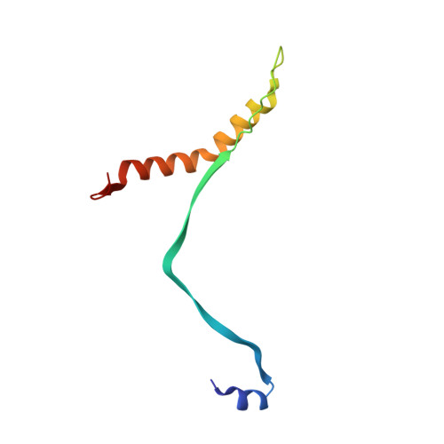
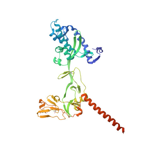
    The paramyxovirus parainfluenza virus 5 (PIV5) enters cells by fusion of the viral envelope with the plasma membrane through the concerted action of the fusion (F) protein and the receptor binding protein hemagglutinin-neuraminidase. The F protein folds initially to form a trimeric metastable prefusion form that is triggered to undergo large-scale irreversible conformational changes to form the trimeric postfusion conformation. It is thought that F refolding couples the energy released with membrane fusion. The F protein is synthesized as a precursor (F0) that must be cleaved by a host protease to form a biologically active molecule, F1,F2. Cleavage of F protein is a prerequisite for fusion and virus infectivity. Cleavage creates a new N terminus on F1 that contains a hydrophobic region, known as the FP, which intercalates target membranes during F protein refolding. The crystal structure of the soluble ectodomain of the uncleaved form of PIV5 F is known; here we report the crystal structure of the cleavage-activated prefusion form of PIV5 F. The structure shows minimal movement of the residues adjacent to the protease cleavage site. Most of the hydrophobic FP residues are buried in the uncleaved F protein, and only F103 at the newly created N terminus becomes more solvent-accessible after cleavage. The conformational freedom of the charged arginine residues that compose the protease recognition site increases on cleavage of F protein.


  • Organizational Affiliation

    Howard Hughes Medical Institute and Department of Molecular Biosciences, Northwestern University, Evanston, IL 60208, USA.


Macromolecules
Find similar proteins by:  (by identity cutoff)  |  3D Structure
Entity ID: 1
MoleculeChains Sequence LengthOrganismDetailsImage
Fusion glycoprotein F2A,
C [auth B],
E [auth C]
81Simian virus 5 (strain W3)Mutation(s): 0 
Gene Names: F
UniProt
Find proteins for P04849 (Parainfluenza virus 5 (strain W3))
Explore P04849 
Go to UniProtKB:  P04849
Entity Groups  
Sequence Clusters30% Identity50% Identity70% Identity90% Identity95% Identity100% Identity
UniProt GroupP04849
Sequence Annotations
Expand
  • Reference Sequence
Find similar proteins by:  (by identity cutoff)  |  3D Structure
Entity ID: 2
MoleculeChains Sequence LengthOrganismDetailsImage
Fusion glycoprotein F1B [auth D],
D [auth E],
F
409Simian virus 5 (strain W3)Mutation(s): 0 
Gene Names: F
UniProt
Find proteins for P04849 (Parainfluenza virus 5 (strain W3))
Explore P04849 
Go to UniProtKB:  P04849
Entity Groups  
Sequence Clusters30% Identity50% Identity70% Identity90% Identity95% Identity100% Identity
UniProt GroupP04849
Sequence Annotations
Expand
  • Reference Sequence
Small Molecules
Ligands 1 Unique
IDChains Name / Formula / InChI Key2D Diagram3D Interactions
NAG
Query on NAG

Download Ideal Coordinates CCD File 
G [auth A]
H [auth D]
I [auth D]
J [auth B]
K [auth E]
G [auth A],
H [auth D],
I [auth D],
J [auth B],
K [auth E],
L [auth E],
M [auth E],
N [auth C],
O [auth C],
P [auth F],
Q [auth F]
2-acetamido-2-deoxy-beta-D-glucopyranose
C8 H15 N O6
OVRNDRQMDRJTHS-FMDGEEDCSA-N
Experimental Data & Validation

Experimental Data

  • Method: X-RAY DIFFRACTION
  • Resolution: 2.00 Å
  • R-Value Free: 0.243 
  • R-Value Work: 0.220 
  • R-Value Observed: 0.221 
  • Space Group: C 1 2 1
Unit Cell:
Length ( Å )Angle ( ˚ )
a = 250.93α = 90
b = 140.52β = 99.84
c = 84.6γ = 90
Software Package:
Software NamePurpose
DENZOdata reduction
SCALEPACKdata scaling
PHASERphasing
PHENIXrefinement
PDB_EXTRACTdata extraction
HKL-2000data collection
HKL-2000data reduction
HKL-2000data scaling

Structure Validation

View Full Validation Report


Ligand Structure Quality Assessment 


Entry History 

Deposition Data

Revision History  (Full details and data files)

  • Version 1.0: 2012-09-19
    Type: Initial release
  • Version 1.1: 2012-10-31
    Changes: Database references
  • Version 1.2: 2017-11-15
    Changes: Advisory, Refinement description
  • Version 1.3: 2020-07-29
    Type: Remediation
    Reason: Carbohydrate remediation
    Changes: Data collection, Database references, Derived calculations, Structure summary
  • Version 1.4: 2023-09-13
    Changes: Data collection, Database references, Refinement description, Structure summary