High-resolution structures of AidH complexes provide insights into a novel catalytic mechanism for N-acyl homoserine lactonase
Gao, A., Mei, G.Y., Liu, S., Wang, P., Tang, Q., Liu, Y.P., Wen, H., An, X.M., Zhang, L.Q., Yan, X.X., Liang, D.C.(2013) Acta Crystallogr D Biol Crystallogr 69: 82-91
- PubMed: 23275166
- DOI: https://doi.org/10.1107/S0907444912042369
- Primary Citation of Related Structures:
4G5X, 4G8B, 4G8C, 4G8D, 4G9E, 4G9G - PubMed Abstract:
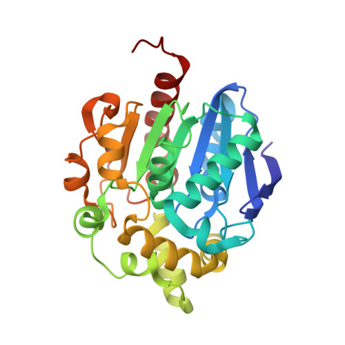
Many pathogenic bacteria that infect humans, animals and plants rely on a quorum-sensing (QS) system to produce virulence factors. N-Acyl homoserine lactones (AHLs) are the best-characterized cell-cell communication signals in QS. The concentration of AHL plays a key role in regulating the virulence-gene expression and essential biological functions of pathogenic bacteria. N-Acyl homoserine lactonases (AHL-lactonases) have important functions in decreasing pathogenicity by degrading AHLs. Here, structures of the AHL-lactonase from Ochrobactrum sp. (AidH) in complex with N-hexanoyl homoserine lactone, N-hexanoyl homoserine and N-butanoyl homoserine are reported. The high-resolution structures together with biochemical analyses reveal convincing details of AHL degradation. No metal ion is bound in the active site, which is different from other AHL-lactonases, which have a dual Lewis acid catalysis mechanism. AidH contains a substrate-binding tunnel between the core domain and the cap domain. The conformation of the tunnel entrance varies with the AHL acyl-chain length, which contributes to the binding promiscuity of AHL molecules in the active site. It also supports the biochemical result that AidH is a broad catalytic spectrum AHL-lactonase. Taken together, the present results reveal the catalytic mechanism of the metal-independent AHL-lactonase, which is a typical acid-base covalent catalysis.
Organizational Affiliation:
National Laboratory of Biomacromolecules, Institute of Biophysics, Chinese Academy of Sciences, Beijing, People's Republic of China.